ADVANTAGES OF OTOENDOSCOPY
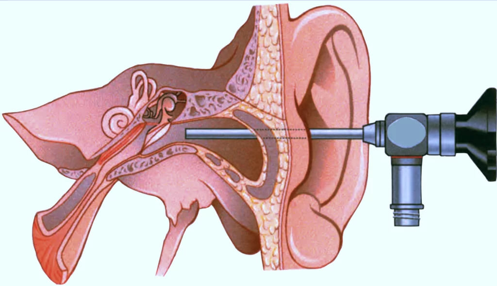
- It provides easy access to sinus tympani, facial recess, epitympanic sinus and protympanum, therefore helps in clearance of disease from these areas. These areas are anatomical blind spots for the operating microscope.
- Manoeuvrability.
- Proximity of image.
- Wide field of vision
- Angle of view
- There is reduced surgical morbidity.
DISADVANTAGES OF OTOENDOSCOPE
- Only one hand free to operate.
- Peri-operative haemorrhage that leads to fogging and smearing of the endoscope tip.
- Limited movement by the extreme curvature of the external auditory canal (EAC).
- Risk of thermal injury to the middle ear
- Unsafe advancement during exploration can cause dislocation or fracture of ossicles.
SPECIFICATIONS OF OTOENDOSCOPE
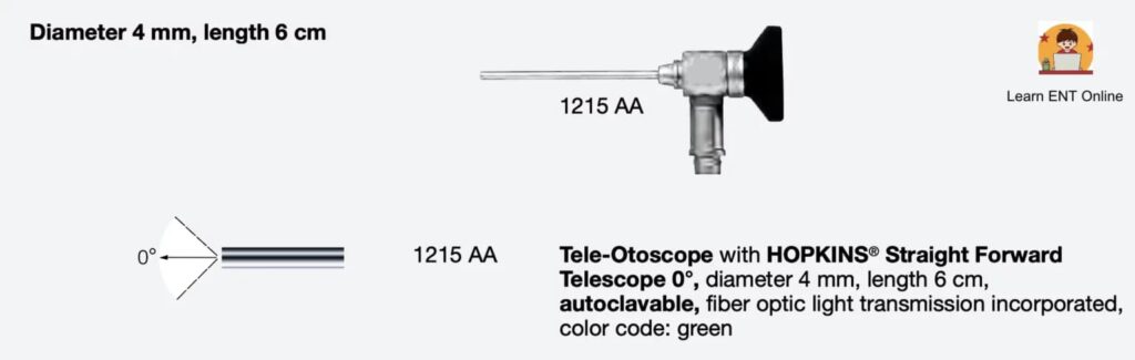
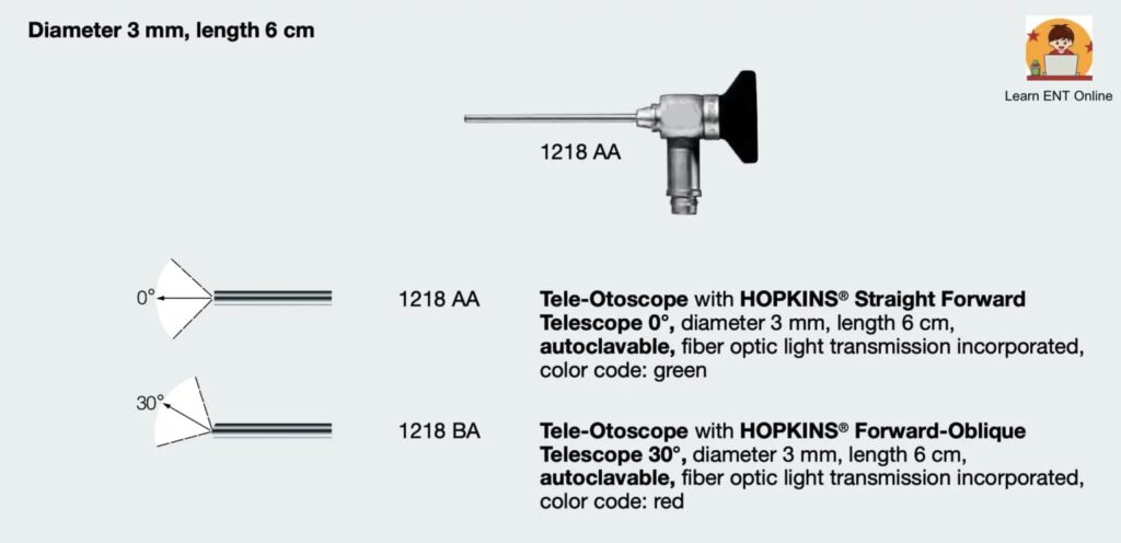
- Generally 3 mm and 4 mm diameter 0, 30 and 45 degree endoscopes are used.
- Rarely, 1.9 mm, 4.0 mm diameter and 70 degree endoscope are also used especially to pass through the posterior tympanotomy.
DIAGNOSTIC INDICATIONS
- Extent of retraction pockets and cholesteatoma. The diagnostic potential of the endoscope lies most commonly in the evaluation of retraction pockets and cholesteatoma, particularly in blind spots such as the sinus tympani, attic, protympanum and hypotympanum.
- Ossicular continuity through perforation. Evaluation of ossicular chain continuity may be undertaken endoscopically through an existing tympanic membrane perforation.
- Otoendoscopy through myringotomy. Middle ear endoscopy may also be carried out through a myringotomy for many indications such as unexplained conductive hearing loss, perilymphatic fistulas or occult cholesteatoma.
- Tympanoplasty/ ossiculoplasty surgery.
- Retraction pocket & cholesteatoma surgery.
- Minimally invasive second look canal wall up mastoid surgery.
- Newer applications include endoscopic stapedotomy, endoscopic cochlear implantation and total transcanal approaches to the inner ear.
Easy access to sinus tympani, facial recess, epitympanic sinus and protympanum, which were the anatomical blind spots for the operating microscope, therefore otoendoscopy helps in clearance of disease from these areas.
———— End of the chapter ————
Download full PDF Link:
Otoendoscopy Best Lecture Notes Dr Rahul Bagla ENT Textbook
Reference Textbooks.
- Scott-Brown, Textbook of Otorhinolaryngology-Head and Neck Surgery.
- Glasscock-Shambaugh, Textbook of Surgery of the Ear.
- P L Dhingra, Textbook of Diseases of Ear, Nose and Throat.
- Hazarika P, Textbook of Ear Nose Throat And Head Neck Surgery Clinical Practical.
- Mohan Bansal, Textbook of Diseases of Ear, Nose and Throat Head and Neck Surgery
- Hans Behrbohm, Textbook of Ear, Nose, and Throat Diseases With Head and Neck Surgery.
- Salah Mansour, Middle Ear Diseases – Advances in Diagnosis and Management.
- Logan Turner, Textbook of Diseases of The Nose, Throat and Ear Head And Neck Surgery.
- Rob and smith, Textbook of Operative surgery.
- Anirban Biswas, Textbook of Clinical Audio-vestibulometry.
- Arnold, U. Ganzer, Textbook of Otorhinolaryngology, Head and Neck Surgery.
Author:

Dr. Rahul Bagla
MBBS (MAMC, Delhi) MS ENT (UCMS, Delhi)
Fellow Rhinoplasty & Facial Plastic Surgery.
Renowned Teaching Faculty
Mail: msrahulbagla@gmail.com
India
———– Follow us on social media ————
- Follow our Facebook page: https://www.facebook.com/Dr.Rahul.Bagla.UCMS
- Follow our Instagram page: https://www.instagram.com/dr.rahulbagla/
- Subscribe to our Youtube channel: https://www.youtube.com/@Drrahulbagla
- Please read. Anatomy of External Ear. https://www.entlecture.com/anatomy-of-ear/
- Please read. Anatomy of Temporal Bone. https://www.entlecture.com/anatomy-of-temporal-bone/
- Please read. Stenger’s, Chimani Moos, Teal test. https://www.entlecture.com/special-tuning-fork-tests/
Keywords: What is otoendoscopy, Otoendoscopy guide for ENT students, Otoendoscopy procedure step by step, Otoendoscopy in ear examination, Diagnostic applications of otoendoscopy, ENT otoendoscopy techniques, Otoendoscopy equipment and uses, Clinical importance of otoendoscopy, Advances in otoendoscopy for ENT, Otoendoscopy vs otoscopy, Otoendoscopy for ear disease diagnosis, How to perform otoendoscopy, Otoendoscopy training for ENT students, Benefits of otoendoscopy in ENT practice, Otoendoscopy common findings, endoscopic ear examination techniques, otoendoscopy equipment reviews, pediatric otoendoscopy procedures, advantages of otoendoscopy in ENT, training courses for otoendoscopy, endoscopic ear surgery recovery time, otoendoscopy for chronic ear infections, comparison of otoendoscopy and otoscopy, latest advancements in otoendoscopy, patient preparation for otoendoscopy.
Excellent academic contribution
Thanks