TRIANGLES OF THE NECK. Anterolaterally, the neck appears as a quadrilateral area. The triangles of the neck help to categorize the anatomy of the neck and in planning neck dissection surgery. Knowledge of these triangles are important because it contains important glands, nerves, vessels and lymph nodes.
| Anterior triangle of neck | – Submental triangle – Submandibular triangle – Carotid triangle – Muscular triangle |
| Posterior triangle of neck | – Occipital triangle – Subclavian triangle |
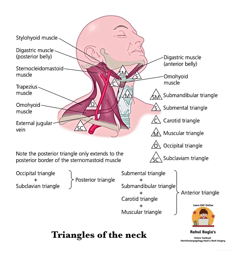
The neck is divided into anterior and posterior triangles by sternocleidomastoid muscle, which passes obliquely from the sternum and clavicle to the mastoid process and occipital bone.
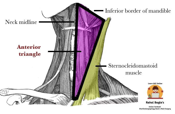
ANTERIOR TRIANGLE OF THE NECK
Boundaries
- Anteriorly – by the midline of the neck.
- Posteriorly – by the anterior border of sternocleidomastoid.
- Superiorly (Base) – by the inferior border of the mandible and a line continued from the angle of the mandible to the mastoid process.
- Apex – by the manubrium sternum.
Contents of Anterior triangle.
| Muscles | Digastric, Mylohyoid, Superior belly of the omohyoid, Stylohyoid and Strap muscles. |
| Vessels | External carotid artery and branches (except posterior auricular), Internal and Anterior jugular vein. |
| Nerves | Internal and External laryngeal nerves, Nerve to mylohyoid, Hypoglossal nerve. |
| Viscera | Thyroid and larynx, Submental and submandibular glands. |
| Other | Jugular chain of lymph nodes. |
Anterior triangle is further divided into submandibular, submental, muscular and carotid triangles by the digastric and omohyoid muscles across the anterior triangle.
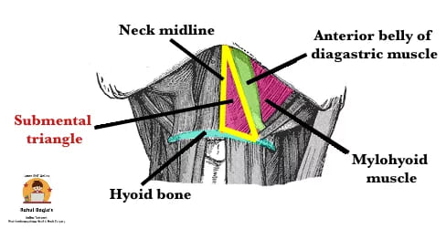
1. Submental triangle. It is a single triangle as compared to the other triangles which are paired on each side of the neck. It is demarcated laterally by the anterior bellies of both digastric muscles. Its apex (superiorly) is formed by symphysis of mandible, its base (inferiorly) is formed by hyoid bone. Its floor is formed by mylohyoid muscle. It contains level Ia lymph nodes 2–8 in number, fatty tissue and small veins which drain into anterior jugular veins.
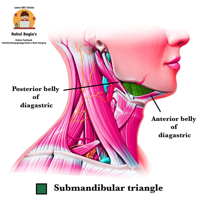
2. Submandibular (Digastric) triangle. It is bounded superiorly by the lower border of body of mandible and its projection to the mastoid process. Posteriorly it is bounded by the posterior belly of digastric and by stylohyoid muscles, and anteriorly by the anterior belly of digastric muscle. Its floor is formed anteriorly by mylohyoid muscle and posteriorly by hyoglossus muscle. It contains the marginal mandibular branch of the facial nerve. It also contains lymph nodes of level Ib and submandibular gland, which has the facial vein superficial to it and the facial artery deep to it. Lower part of the parotid gland is present in posterior region of the submandibular triangle.
3. Muscular triangle. It is bounded anteriorly by midline from the hyoid bone to the sternum. Inferolaterally by the anterior border of sternocleidomastoid and superolaterally by the superior belly of omohyoid. The triangle contains strap muscles, parathyroid glands and the upper aerodigestive tract.
4. Carotid triangle. It is bounded laterally by posterior border of sternocleidomastoid, medially by the superior belly of omohyoid and superiorly by the posterior belly of digastric. It floor is formed by thyrohyoid, hyoglossus muscles and inferior and middle pharyngeal constrictor muscles. It contains the arteries (common carotid artery and its division into external and internal carotid arteries), nerves (vagus nerve, the hypoglossal nerve, the superior root of the ansa cervicalis, the superior laryngeal nerve) and lymph nodes of the jugular chain.
.
POSTERIOR TRIANGLE OF THE NECK
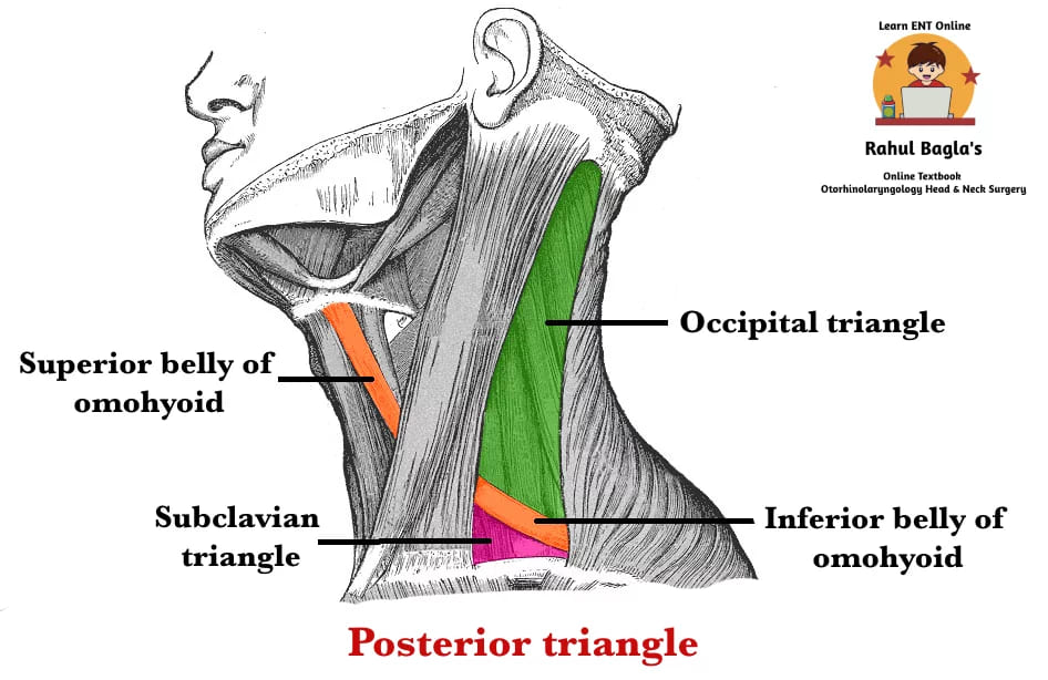
Boundaries
- Anteriorly by the posterior border of sternocleidomastoid.
- Posteriorly by the anterior border of trapezius.
- Inferiorly by the clavicle.
- Apex is between the attachments of sternocleidomastoid and trapezius to the occiput.
- Roof is formed by the investing layer of the deep cervical fascia.
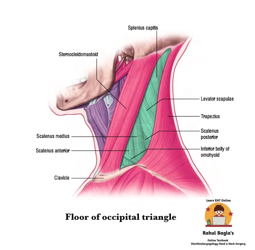
- Floor is formed by the prevertebral fascia covering splenius capitis, levator scapulae and the scalene muscles.
Contents of Posterior triangle.
| Muscles | Omohyoid |
| Vessels | Occipital, transverse cervical, suprascapular and subclavian arteries, Transverse cervical, suprascapular and external jugular veins. |
| Nerves | Spinal accessory nerve, Cervical and brachial plexus |
| Other | It contains level V lymph nodes (include lymph nodes of spinal accessory chain, transverse cervical nodes and supraclavicular nodes) |
Posterior triangle is divided into occipital and supraclavicular triangles by the inferior belly of omohyoid, which crosses approximately 2.5 cm above the clavicle.
1. Occipital triangle. It is the upper and bigger part of the posterior triangle. Anteriorly it is bounded by the posterior border of sternocleidomastoid. Posteriorly by the anterior border of trapezius. Inferiorly it is bounded by the inferior belly of omohyoid. Its floor is formed by splenius capitis, levator scapulae, and scaleni muscles (anterior, medius and semispinalis capitis occasionally.
The triangle is covered by skin, superficial and deep fasciae, and inferiorly by platysma. It contains spinal accessory nerve (CN XI), branches of the cervical plexus, upper most part of brachial plexus, supraclavicular nerve.
2. Subclavian triangle. It is the lower and smaller part of the posterior triangle. The boundaries of the subclavian triangle are the inferior belly of the omohyoid, the clavicle and the posterior border of the sternocleidomastoid muscle. Its contains the branchial plexus, supraclavicular nerves, the third part of the subclavian artery, first rib, scalenus medius and the first slip of serratus anterior. The domes of the pleura with the overlying supra-pleural fascia is also present in this triangle.
——– End of the chapter ——–
Reference Textbooks.
- Scott-Brown, Textbook of Otorhinolaryngology-Head and Neck Surgery.
- Cummings, Otolaryngology-Head and Neck Surgery.
- Stell and Maran’s, Textbook of Head and Neck Surgery and Oncology.
- Ballenger’s, Otorhinolaryngology Head And Neck Surgery
- Susan Standring, Gray’s Anatomy.
- Frank H. Netter, Atlas of Human Anatomy.
- B.D. Chaurasiya, Human Anatomy.
- P L Dhingra, Textbook of Diseases of Ear, Nose and Throat.
- Hazarika P, Textbook of Ear Nose Throat And Head Neck Surgery Clinical Practical.
- Mohan Bansal, Textbook of Diseases of Ear, Nose and Throat Head and Neck Surgery.
- Hans Behrbohm, Textbook of Ear, Nose, and Throat Diseases With Head and Neck Surgery.
- Logan Turner, Textbook of Diseases of The Nose, Throat and Ear Head And Neck Surgery.
- Arnold, U. Ganzer, Textbook of Otorhinolaryngology, Head and Neck Surgery.
- Ganong’s Review of Medical Physiology.
Author:

Dr. Rahul Bagla
MBBS (MAMC, Delhi) MS ENT (UCMS, Delhi)
Fellow Rhinoplasty & Facial Plastic Surgery.
Renowned Teaching Faculty
Mail: msrahulbagla@gmail.com
India
Please read. Glomus Tumour. https://www.entlecture.com/glomus-tumour/
Follow our Facebook page: https://www.facebook.com/Dr.Rahul.Bagla.UCMS
Join our Facebook group:
Hello. Im a new ENT resident. Just found this website 2 weeks ago and I Iove it.
It’s easy to read and understand.
Key points are highlighted.
I really do appreciate the effort that was put into making this website
Thank you so much for your kind words