Physiology of Hearing. Hearing is defined as the neural perception of sound energy. It involves the identification (what?) and localisation (from where?) of the sounds. An ear is a statoacoustic organ. The external ear, middle ear and cochlea of the inner ear are involved in the mechanism of hearing while the semicircular canals, utricle and saccule of the inner ear maintain equilibrium (balance).
What is Sound ?
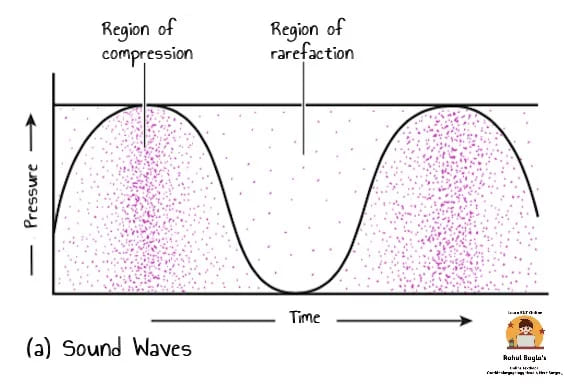
Sound waves are caused by the compression and decompression of air molecules by any vibrating object.
Formation of sound waves:
(a) Sound waves are alternating regions of compression and rarefaction of the air molecules.
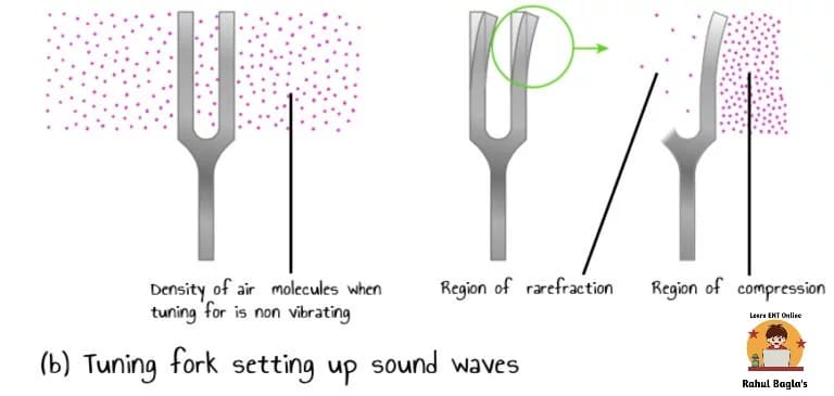
(b) A vibrating tuning fork sets up sound waves, as the air molecules ahead of the advancing arm of the tuning fork are compressed while the molecules behind the arm are rarefied.
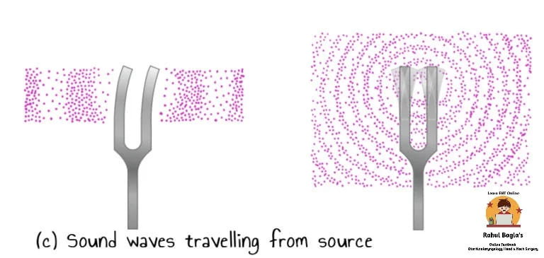
(c) Disturbed air molecules bump into molecules beyond them, setting up new regions of air disturbance more distant from the original source of the sound. In this way, sound waves travel progressively farther from the source, even though each air molecule travels only a short distance when it is disturbed. The sound wave dies out when the last region of air disturbance is too weak to disturb the region beyond it.
Speed of sound:
- In the air, at 20°C and at sea level, sound travels at a speed of 344 m (1120 ft) per second. It travels faster in liquids and solids than in the air.
- When sound waves passes from air to liquid medium, it travel less efficiently, because most of sound waves are reflected due to high inertia offered by the liquid.
So the question arises, How sound reaches the brain and what is its mechanism ?
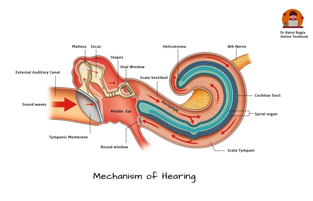
- The sound waves in the environment are localized and captured by the pinna and directs the sound waves into the external auditory meatus.
- Then the sound waves crosses through external auditory canal and strikes the tympanic membrane and causes vibrations in the tympanic membrane.
- These vibrations are then transmitted to stapedovestibular joint through malleus and incus. Movements of stapedovestibular joint causes pressure changes in the labyrinthine fluids.
- Movement of labyrinthine fluids causes movement of the basilar membrane. Movement in the basilar membrane causes stimulation of the hair cells which are present on it.
- Hair cells (also known as transducers) which are situated in the Rosenthal’s canal (canal running along osseous spiral lamina) converts this mechanical energy into electrical energy (transduction process) and transfer these electrical impulses to the auditory nerve.
Auditory Pathway:
- Hair cells are innervated by dendrites of bipolar cells of spiral ganglion.
- Central axons of spiral ganglion bipolar cells collect to form the cochlear division of Eighth cranial nerve.
- The cochlear nerve ends in the Cochlear nuclei (both dorsal and ventral) in the medulla.
- From here, both crossed and uncrossed fibres travel to the superior Olivary nucleus, Lateral lemniscus, Inferior colliculus, Medial geniculate body and finally reach the Auditory cortex of the temporal lobe (Brodmann’s area 41 situated at the superior aspect of the temporal lobe along the floor of the lateral cerebral fissure), located mainly in the superior gyrus of the temporal lobe.
- Other important areas in the temporal lobe are: Primary auditory cortex (areas 42) and Auditory association areas (areas 22, 21 and 20).
To remember auditory pathway, remember the mnemonic E COLI-MA (Eight nerve, cochlear nucleus, olivary complex, lateral lemniscus, inferior colliculus Medial geniculate body, Auditory cortex).
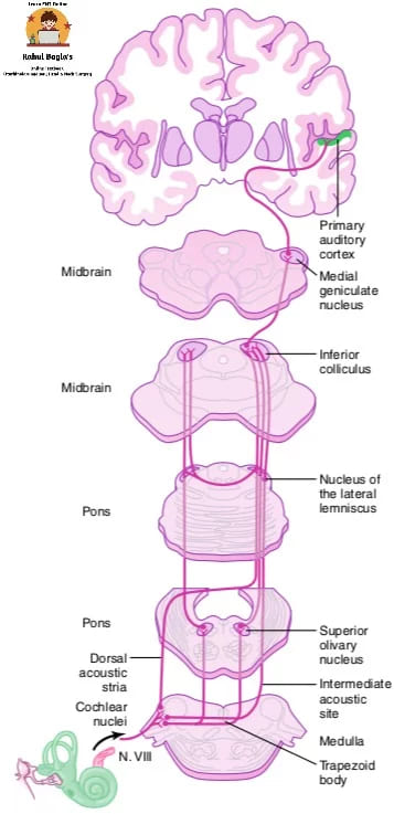 Auditory pathways from the right cochlea. Note bilateral route through brainstem and bilateral cortical representation.
Therefore, the mechanism of hearing can be broadly divided into:
- Mechanical conduction of sound (conduction apparatus).
- Transduction of mechanical energy to electrical impulses (sensory system of cochlea).
- Conduction of electrical impulses to the brain (neural pathways).
1. Mechanical conduction of sound (conduction apparatus).
Role of External Ear:
(i) Pinna: Localizes, captures and directs the sound into the external auditory meatus.
(ii) External auditory meatus: Conducts the sound waves to the tympanic membrane leading to its vibration.
(iii) Tympanic membrane: The vibrations of the tympanic membrane are transmitted via the chain of ossicles to the stapes footplate. Thus, the tympanic membrane acts as:
(a). Pressure receiver, i.e. it is extremely sensitive to pressure changes produced by the sound waves.
(b). Resonator, i.e. starts vibrating with pressure changes produced by the sound waves.
(c). Critically dampens, i.e. the vibrations of tympanic membrane cease immediately after the end of the sound.
Role of Middle ear: There are four mechanism through which middle ear plays important role in hearing mechanism.
(i) Impedance matching mechanism (also known as Transformation action)
(ii) Attenuation reflex.
(iii) Phase differential between cochlear windows.
(iv) The natural resonance of external ear and middle ear.
(i) Impedance matching mechanism. The vibration transfer is not as simple as it seems because the acoustic impedance (resistance) of fluid in the inner ear is much more than the air in the middle ear (i.e., impedance mismatch).
Hence, greater force is required to cause vibration in the fluid. 99.9% of sound energy is reflected away from the surface of the water when sound travels from air into water. To compensate this loss, the tympanic membrane and the ear ossicles together convert the sound of greater amplitude but lesser force, to a sound of lesser amplitude but greater force. This function is called as “ impedance matching mechanism or the transformer action.”.
It is accomplished by following three mechanisms:
- The lever action of ossicles. The handle of malleus is 1.3 times longer than the long process of incus, this provides a mechanical leverage advantage, due to which the middle ear ossicles increase the force of movement by 1.3 times.
- The hydraulic action of the tympanic membrane. The surface area of the tympanic membrane is much larger than the surface area of stapes footplate. The average ratio between the two being 21:1. As the effective vibratory area of the tympanic membrane is only two-thirds of the total surface area. The effective areal ratio is reduced to 14:1. This size differences between the tympanic membrane and stapes footplate provides the mechanical advantage. It means the force produced by the sound is concentrated over a smaller area, thus amplifying the pressure exerted on the oval window. Therefore, the product of areal ratio and lever action of ossicles is 18:1 (i.e., 18 folds). Detailed calculation is given below in the diagram.
- Curved tympanic membrane effect. Movements of the tympanic membrane are more at the periphery than at the centre where malleus handle is attached. This too provides some leverage.
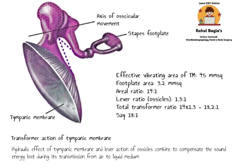
Another study (Wever and Lawrence) concluded that out of a total of 90 mm2 area of the human tympanic membrane, only 55 mm sq is functional and given the area of stapes footplate (3.2 mm sq), the areal ratio is 17:1 and total transformer ratio (17× 1.3) is 22.1.
(ii) Attenuation reflex (acoustic reflex) is a protective reflex. The muscles in the middle ear contract in response to ambient sounds >70-80 dB above hearing threshold, thereby tightening the tympanic membrane and restricting the movement of the ossicular chain. Hence the transmission of intense sound to the inner ear is stopped to protect the delicate sensory apparatus from damage.
Mechanism: Contraction of tensor tympani muscle pulls the malleus inwards whereas contraction of stapedius muscle pulls stapes outwards. These two opposing forces make the ossicular system very rigid and sound is not allowed to enter the inner ear. Contractions of the tensor tympani have also been associated with the opening of the eustachian tube where the inward motion of the tympanic membrane that results from the contraction produces an overpressure in the middle ear that helps open the tube.
Advantages:
- It prevents cochlear damage from loud music, the sound of jet aircraft, etc.
- It masks the low-frequency environmental sounds and allows us to concentrate on the sound above 1000 Hz.
- It reduces the intensity of the sounds occurring just prior to vocalisation and chewing.
Note: The attenuation reflex is a slow reflex with a latent period of 40 milliseconds and it occurs with a slight delay after extremely loud sound (gun-shot or bomb explosion). This slight delay may cause deafness due to cochlear damage. It thus provides protection only from prolonged intense sounds, not from sudden sounds like an explosion. So for this reason during World War II anti-aircraft guns were designed to make a loud pre-firing sound to protect the gunner’s ears from the much louder sound of the actual firing.
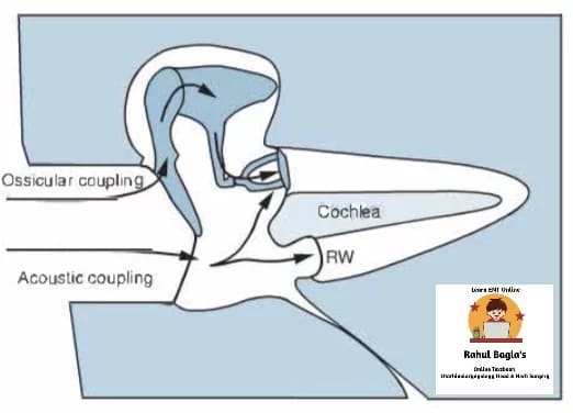
(iii) Phase differential between cochlear windows. Normally, the fluids in the inner ear are incompressible. The round window vibrates in opposite phase to the oval window, in doing so it allows fluid in the cochlea to move. It means that when the oval window receives a wave of compression, the round window is at the phase of rarefaction. Thus, if the sound waves were to strike both the windows at the same time and same phase, they would cancel each other effect and there will be no movement of perilymph and no hearing. Phase differential between the windows contributes 4 dB when the tympano-ossicular system is intact. This acoustic separation of windows is attributed to the
- “Ossicular coupling” effect is produced by an intact tympano-ossicular system (preferential pathway to the oval window).
- “Acoustic coupling” effect is produced by a cushion of air in the middle ear around the round window also leads to phase difference.
(iv) Natural resonance of external and middle ear. The external ear and middle ear, due to the inherent anatomic and physiologic properties, allows certain frequencies of sound to pass more easily to the inner ear. The natural resonance of important structures is:
- External auditory canal is 3000 Hz
- Tympanic membrane is 800–1600 Hz
- Middle ear is 800 Hz
- Ossicular chain is 500–2000 Hz
Thus, the greatest sensitivity of the sound transmission is between 500 and 3000 Hz, and used in daily talk and conversation.
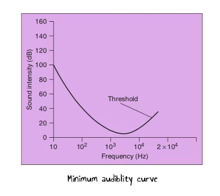
Minimum audibility curve:
- The amplification of sound intensity is highest between 1000 and 3000 Hz.
- The sounds below 16Hz or above 20,000Hz are never amplified.
- This is the reason, why human ear can perceive the pitch of sound between 16 and 20,000 Hz. Maximum sensitivity is between 1000 and 3000 Hz. This effect is depicted in the minimum audibility curve
2. Transduction of mechanical energy to electrical impulses (sensory system of the cochlea).
The conversion of mechanical energy to electric nerve impulse occurring in the hair cells of the organ of Corti is termed ‘auditory transduction’.
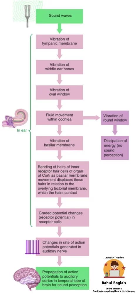 Auditory transduction pathway
Process: Stapes footplate is attached to the oval window which is in close contact with scala vestibuli (containing perilymph). Movements of stapes footplate bring pressure changes in the perilymph which are transmitted to reissner’s membrane, causing compression of scala media (containing endolymph).
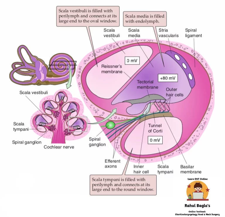
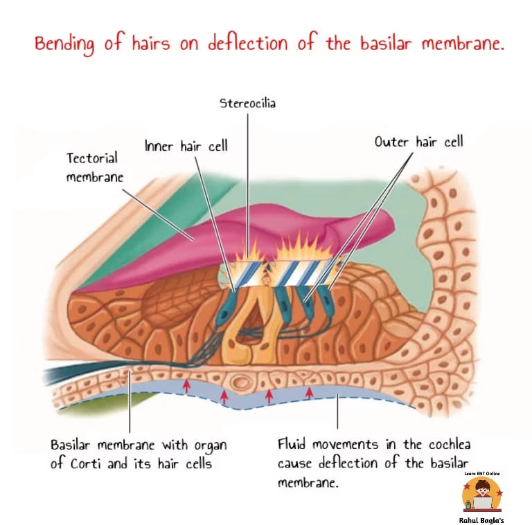
The pressure wave in endolymph causes relative deflection of the basilar membrane in relation to the overlying stationary tectorial membrane; as a result of which hearing forces develop, distorting and bending the stereocilia (hairs).
The bending of stereocilia open mechanically-gated cation channels leading to ion movement (K+ and Ca+) and receptor potential.
- When the organ of Corti moves up, the tectorial membrane slides forward relative to the basilar membrane, bending the stereocilia away from the limbus causing depolarization (RMP is -60mV and depolarization -50mV).
- When the organ of Corti moves down, the tectorial membrane slides backwards relative to the basilar membrane and bends the stereocilia towards the limbus causing hyper-polarization.
Electrical response of hair cells. Using microelectrodes, four gross potentials are recorded from the ear:
(i) Endo-cochlear potential: It is a direct current potential (+80 mv) recorded from scala media. It is present in resting conditions i.e in absence of any external auditory stimulus. This potential provides a source of energy for cochlear transduction
Source: Endolymph contains more K+(150 mEq/litres) than perilymph (3 mEq/litres) due to the presence of Na+/K+-ATPase pump in stria vascularis of scala media.
(ii) Cochlear microphonic potential: An alternating current is recorded when the auditory stimulus is presented to the ear. This is the sum of receptor potentials of a large number of hair cells when recorded extracellularly. One electrode is placed on scala media and another in scala tympani.
Source: When basilar membrane moves in response to a sound stimulus, electrical resistance at the tips of hair cells changes allowing the flow of K+ through hair cells and produces voltage fluctuations called cochlear microphonics.
(iii) Summating potential (SP): It is a direct current potential superimposed on the auditory nerve action potential. SP is used for diagnosis of Meniere’s disease. Both Cochlear Microphonics and Summating Potentials are receptor potentials as seen in other sensory end-organs. They differ from action potentials in that:
- Both are graded potential and not, action potential
- Are not propagated
- Have no post-response refractory period.
- Have no latency
(iv) Compound action potential: It is an all or none response of auditory nerve fibres.
3. Conduction of electrical impulses to the brain (neural pathways).
Neural transmission of signals – Auditory pathways comprise the following relay stations:
(i) Spiral ganglion (First-order neurons). First-order neurons are the bipolar cells of the spiral ganglion, which is situated in the Rosenthal’s canal (canal running along osseous spiral lamina). Hair cells get innervation from dendrites of bipolar cells of spiral ganglion, Axons of which forms the cochlear division of eighth cranial nerve. The cochlear nerve ends in the cochlear nuclei in the upper part of the medulla.
(ii) Superior olivary nucleus complex, trapezoid nucleus and nucleus of lateral lemniscus (Second-order neurons). Second-order neuronshave their cell bodies in the cochlear nuclei, located in the medulla. There are two cochlear nuclei: dorsal and ventral. At this point, all the fibres synapse, and second-order neurons pass mainly to the opposite side of the brain stem to terminate in the superior olivary nucleus. A few second-order fibres also pass to the superior olivary nucleus on the same side.
(iii) Inferior colliculus (Third-order neurons). Third-order neuronshave their cell bodies mainly in the superior olivary complex (made up of a number of nuclei) and also in trapezoid nucleus and nucleus of the lateral lemniscus. From the superior olivary nucleus, the auditory pathway passes upward through the lateral lemniscus. Some of the fibres terminate in the nucleus of the lateral lemniscus, but many bypass this nucleus and travel on to the inferior colliculus, where all or almost all the auditory fibres synapse.
- Superior olivary nuclear complex receives the large majority of lateral lemniscus fibres from the cochlear nuclei. Axons arising from the superior olivary complex form an important ascending bundle called the lateral lemniscus.
- Trapezoid nucleus. Some cochlear fibres that do not relay in the superior olivary nucleus join the lateral lemniscus after relaying in the scattered groups of cells lying within the trapezoid body (which constitute the trapezoid nucleus).
- Nucleus of lateral lemniscus refers to the collection of cells that lie within the lemniscus itself. Some cochlear fibres relay in these cells. The fibres of lateral lemniscus ascend to the midbrain and terminate in the inferior colliculus.
(iv) Medial geniculate body (Fourth-order neurons). Fourth-order neuronshave their cell bodies in the inferior colliculus, where the fibres of lateral lemniscus terminate. Fibres arising in the inferior colliculus enter the inferior brachium to reach the medial geniculate body (Fifth-order neurons) where all the fibres do synapse. Fibres arising in the medial geniculate body form
(v) Auditory cortex. (Fifth-order neurons). Finally, the pathway proceeds by way of the auditory radiation to the auditory cortex (Brodmann’s area 41 situated at the superior aspect of the temporal lobe along the floor of the lateral cerebral fissure), located mainly in the superior gyrus of the temporal lobe. Other major areas constituting auditory cortex present in the temporal lobe are: Primary auditory cortex (areas 42) and Auditory association areas (areas 22, 21 and 20).
Neural processing of auditory information involves:
Encoding of sound frequency: The human auditory mechanism can discriminate between the sounds in the range of 60-20,000 Hz. Cochlear nerve fibres encode the frequency of the sound stimulus. Duplex theory( including both, place theory and frequency theory) explains the frequency coding of sound.
Encoding of intensity: occurs at the level of cochlear nerve fibres by the following mechanisms:
- With the increase in the intensity of sound wave, the amplitude of vibration of the basilar membrane increases, which in turn increases the frequency of firing in an auditory nerve fibre.
- As the amplitude of vibration increases, a major portion of the basilar membrane is vibrated, the hair cells are stimulated and auditory nerve fibres are activated.
- Certain inner hair cells are stimulated by very loud sound & they apprise the nervous system that intensity of sound is high.
Feature detection: Higher auditory centres respond to particular features of sound stimuli. For example, cortical neurons may respond specifically to a shift from high-to-low-frequency notes, which is why lesions of the auditory cortex may not impair the ability to discriminate frequency. Instead, lesions of auditory cortex cause a loss of ability to recognise a patterned sequence of sounds.
Sound location in space: A human can distinguish sounds originating from the sources separated by as little as 1°. The binaural receptive field is a feature of most auditory neurons above the level of cochlear nuclei and these contribute to sound localization. The auditory system uses the time lag between the entry of sound in the two ears as a clue to judge the origin of the sound. The inter-aural time difference even if it is as little as 20 μs, is an important clue, especially for low-frequency sounds (below 3000 Hz). However, sound localization is markedly disrupted in the lesions of the auditory cortex.
Physiology of Hearing Physiology of Hearing PPhysiology of Hearing Physiology of Hearing Physiology of Hearing Physiology of Hearing Physiology of Hearing Physiology of Hearing Physiology of Hearing Physiology of Hearing Physiology of Hearing Physiology of Hearing Physiology of Hearing Physiology of Hearing Physiology of Hearing Physiology of Hearing Physiology of Hearing Physiology of Hearing Physiology of Hearing Physiology of Hearing hysiology of Hearing Physiology of Hearing Physiology of HePhysiology of Hearing Physiology of Hearing Physiology of Hearing Physiology of Hearing Physiology of Hearing Physiology of Hearing Physiology of Hearing aring
OTHER IMPORTANT TERMS:
- Amplitude (intensity) determines the loudness of the sound and is measured in decibels.
- Frequency refers to the number of waves produced per second. The unit of frequency is hertz (Hz). Range of human hearing is approximately 20–20,000 Hz. The average range of normal speech is 2000–5000 Hz.
- Pitch. It is the subjective sensation produced by the frequency of sound. Higher the frequency greater is the pitch. Pitch of the average male voice is 120 Hz and that of a female is 250 Hz. An average individual can distinguish about 2000 different pitches.
- Pure Tone. A single frequency sound is called a pure tone, e.g. a sound of 250, 500 or 1000 Hz. In pure tone audiometry, we measure the threshold of hearing in decibels for various pure tones from 125 to 8000 Hz.
- Complex Sound refers to more than one frequency. For example, a human voice is a complex sound.
- Sound pressure is expressed in decibels (dB), which is a relative measure on a log scale.
- Overtones. A complex sound is a mixture of pure tones. The lowest frequency at which a source vibrates is called fundamental (or primary) frequency. All other frequencies, which are multiples of the fundamental frequency are called overtones or harmonics. The overtones determine the quality or the timbre of the sound. Variations in timbre permit us to identify the sounds of various musical instruments (e.g. guitar, piano, tabla, sarangi etc.) even though they are playing notes of the same pitch.
Please read. Hearing tests. Click on this link. https://www.entlecture.com/assessment-of-hearing/
Like our Facebook page: https://www.facebook.com/Dr.Rahul.Bagla.UCMS
Join our Facebook group: https://www.facebook.com/groups/628414274439500
———— End of the chapter ————
Learning resources.
- Scott-Brown, Textbook of Otorhinolaryngology Head and Neck Surgery.
- Ganong’s Review of Medical Physiology.
- Indu Khurana, Textbook of Medical Physiology
- Lauralee Sherwood, Human Physiology: From Cells to Systems
- Glasscock-Shambaugh, Textbook of Surgery of the Ear.
- Logan Turner, Textbook of Diseases of The Nose, Throat and Ear Head And Neck Surgery.
- P L Dhingra, Textbook of Diseases of Ear, Nose and Throat.
- Hazarika P, Textbook of Ear Nose Throat And Head Neck Surgery Clinical Practical.
- Mohan Bansal, Textbook of Diseases of Ear, Nose and Throat Head and Neck surgery.
- Anirban Biswas, Textbook of Clinical Audio-vestibulometry.
Author:

Dr. Rahul Kumar Bagla
MS & Fellow Rhinoplasty & Facial Plastic Surgery.
Associate Professor & Head
GIMS, Greater Noida, India
msrahulbagla@gmail.com
