ANATOMY OF THE EXTERNAL NOSE
The External nose is the first part of the upper respiratory tract and is responsible for warming, humidifying and filtering of inspired air. It contains the peripheral organ of smell. The external nose opens anteriorly onto the face through the nostrils or nares. It is a pyramidal projection in the mid face with its root up and the base directed downwards. The nasal pyramid consists of an osteocartilaginous framework covered by muscles and skin.
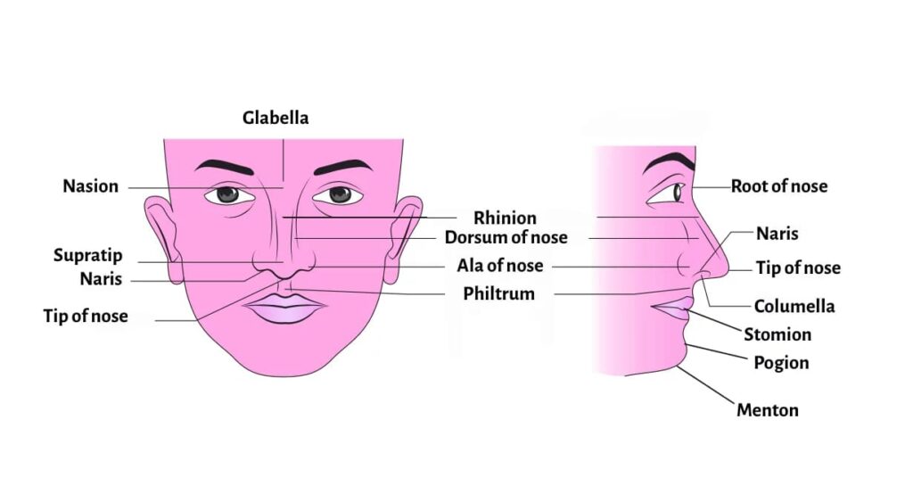
It presents the following features:
- Tip of the nose. It is the lower free end of the nose
- Supratip. It is present just above the tip of the nose.
- Root or bridge. It is the uppermost narrow part of the nose.
- Nasion. It is the junction between the frontal bones and the nasal bones.
- Rhinion. It is the junction between the nasal bones and cartilages.
- Dorsum.
- Nostrils or nares.
- Ala. It is formed by alar cartilages.
Osteocartilaginous Framework (The Skeleton)
The skeletal framework of the nose determines its shape and structural integrity. Hence, understanding the bony and cartilaginous components is crucial for fracture management and rhinoplasty.
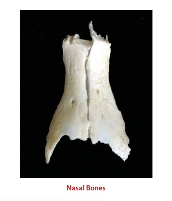
1. Bony Part (Upper One-Third)
The upper one-third of the external nose is bony, while the lower two-thirds are cartilaginous. The bony part consists of two small rectangular nasal bones that meet in the midline. It forms the bridge of the nose.
Boundaries of nasal bones:
- Superior Border: Articulates with the nasal process of the frontal bone.
- Inferior Border: Is related to the upper lateral cartilage (at the Rhinion).
- Lateral Border: Articulates with the frontal process of the maxilla.
- Medial Border: Articulates with the opposite nasal bone and forms a small part of the nasal septum.
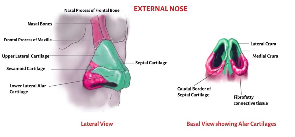
2. Cartilaginous Part (Lower One-Third)
The nasal cartilages are hyaline cartilages that are attached to the bones to form the skeletal framework of the external nose.
It consists of:
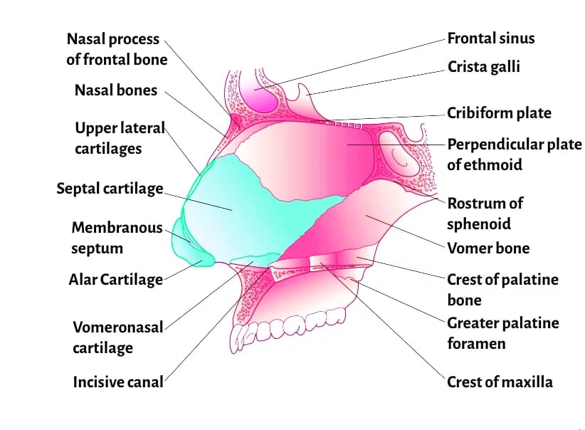
- Upper lateral cartilages. They are trapezoid-shaped and extend from the undersurface of the nasal bones (rhinion) above to the lower lateral cartilages and alar cartilages below. They fuse with each other and attach to the upper border of the midline septal cartilage. The lower free edge of the upper lateral cartilage is seen intranasally as limen vestibule, nasal valve or limen nasi on each side.
- Lower lateral cartilages (alar cartilages). Each alar cartilage is U-shaped. It has a lateral crus, which forms the ala and a medial crus, which runs in the columella and forms the natural arch of the nasal ala. The lateral crus overlaps the lower edge of the upper lateral cartilage on each side.
- Lesser alar (or sesamoid) cartilages. Two or more in number. They lie above and lateral to the alar cartilages. The various cartilages are connected with one another and with the adjoining bones by perichondrium and periosteum. Most of the free margin of the nostril is formed of fibrofatty tissue and not the alar cartilage.
- Septal cartilage. Its anterosuperior border runs from under the nasal bones to the nasal tip. It supports the dorsum of the cartilaginous part of the nose. In a septal abscess or after excessive removal of septal cartilage, as in a submucosal resection (SMR) operation, support of the nasal dorsum is lost and a supratip depression results.
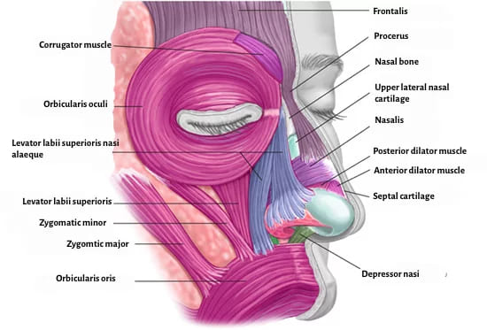
Nasal muscles (Facial Expression and Function)
The osteocartilaginous framework of the nose is covered by muscles that function to elevate, depress, compress, or dilate the nostrils and nasal tip.
- Nasal elevator muscles (Pull nose up) – Procerus, Levator labii-superioris alaeque nasi, Anomalous nasi muscles.
- Nasal depressor muscles (Pull tip down) – Alar nasalis and Depressor septi nasi.
- Compressor muscles (Close nostril) – Transverse nasalis.
- Dilator muscles (Open nostril) – Dilator naris anterior.
Nasal skin
The skin over the nasal bones and upper lateral cartilages is thin and loosely adherent to the underlying framework. The skin over the nasal tip and alar cartilages becomes thicker and more adherent, and contains numerous sebaceous glands. It is the hypertrophy of these sebaceous glands that gives rise to a lobulated tumour called rhinophyma.
Vestibule of the nose
It is the most anterior and inferior aspect of the nasal cavity. It is also the entry point from the external nose into the nasal cavity. It is lined by skin and contains sebaceous glands, sweat glands and coarse hairs called vibrissae. Vibrissae filter the airborne particles during inspiration. The caudal border of the lower lateral cartilage on the lateral wall demarcates the limen nasi (also called the nasal valve), which is the upper limit of the vestibule. Incision is given in the limen nasi during external approach rhinoplasty.
Nasal Valve Area
The Nasal Valve Area is the least cross-sectional area of the entire nose. Therefore, it is the most critical area for regulating airflow and nasal resistance on inspiration. The angle between the nasal septum and the lower border of the upper lateral cartilage is nearly 30° (a few sources say 10° to 15°). When this angle narrows (e.g., after trauma or rhinoplasty), it causes significant nasal obstruction.
Nasal Valve Boundaries:
- Laterally: The lower border of the Upper Lateral Cartilage and fibrofatty tissue (along with the anterior head of the inferior turbinate).
- Medially: The cartilaginous nasal septum.
- Caudally (Floor): The floor of the pyriform aperture.
Blood supply of the external nose
Arterial supply – Branches of both external and internal carotid artery supplies the external nose.
- Nasal dorsum and tip – Lateral nasal branch of the Angular Artery (External Carotid System) & External nasal branch of the Anterior Ethmoid Artery (Internal Carotid System)
- Nasal side wall – External nasal branch of the infraorbital artery.
- Alar region – Angular and superior labial arteries.
- Columella – Columellar branch of the superior labial artery

Venous drainage – Drains into the facial vein and ophthalmic vein. Drainage into the Ophthalmic Vein connects the nose to the Cavernous Sinus via the deep facial vein. Therefore, infections of the nasal tip or ala (known as the “Danger Area” of the face) can potentially lead to Cavernous Sinus Thrombosis.
Lymphatic drainage – It drains into the submandibular, submental and facial nodes.
Nerve supply of the external nose – It is clinically important for the surgeon while giving nerve block for procedures such as rhinoplasty or closed reduction of nasal fractures.
- Supra and infratrochlear branches of the ophthalmic nerve (V1) – Root of the nose, upper part of the nasal bridge and upper part of the lateral wall of the nose.
- Anterior ethmoid nerve branch of the ophthalmic nerve (V1) – Lower part of the nasal bridge and the nasal tip.
- Infraorbital branch of the maxillary nerve (V2) – Lower part of the lateral wall of the nose.
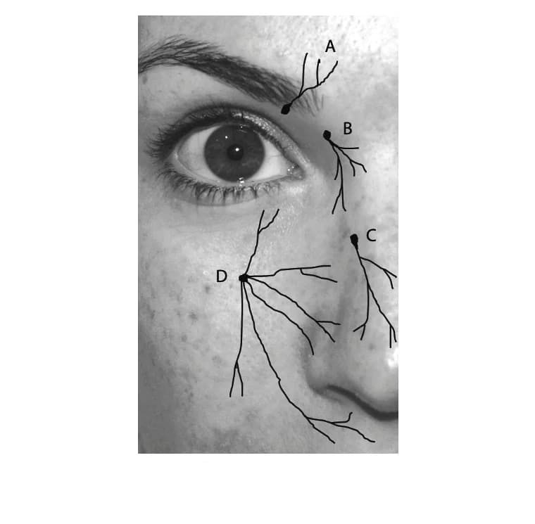
(A) supratrochlear nerve (B), infratrochlear nerve (C), anterior ethmoid nerve (D), and infraorbital nerve.
———–End———–
Multiple Choice Questions (MCQs) (NEET PG Style)
- Which structure forms the anatomical junction known as the Rhinion? A. Frontal bone and Nasal bone B. Nasal bone and Upper lateral cartilage C. Upper lateral cartilage and Septal cartilage D. Lower lateral cartilage and Alar cartilage
- The most crucial area for regulating nasal airflow resistance on inspiration is: A. The Nares B. The Nasal Dorsum C. The Pharyngeal Isthmus D. The Nasal Valve Area
- Infection of the nasal tip can pose a risk of spreading intracranially due to venous drainage into which of the following? A. Jugular vein B. Ophthalmic vein C. Superior sagittal sinus D. Internal maxillary vein
- A patient develops a lobulated, bulbous nose due to severe hypertrophy of sebaceous glands. This condition is termed: A. Rhinitis Sicca B. Saddle Nose Deformity C. Rhinophyma D. Antrochoanal Polyp
- Which cartilage’s excessive removal during a Septoplasty operation leads to a loss of dorsal support and a supratip depression? A. Lower lateral cartilage B. Upper lateral cartilage C. Septal cartilage D. Lesser alar cartilage
- The medial boundary of the Nasal Valve Area is formed by the: A. Inferior turbinate B. Cartilaginous nasal septum C. Lateral crus of the lower lateral cartilage D. Fibrofatty tissue
- The main sensory nerve supply to the Nasal Tip is a branch of the Ophthalmic Nerve (V1) known as the: A. Infraorbital nerve B. Supratrochlear nerve C. Anterior ethmoid nerve D. Maxillary nerve
- Which border of the nasal bone articulates with the frontal process of the maxilla? A. Superior border B. Inferior border C. Medial border D. Lateral border
- The coarse hairs in the vestibule of the nose, responsible for initial filtering of inspired air, are called: A. Cilia B. Microvilli C. Vibrissae D. Turbinates
- The arterial supply to the nasal dorsum includes the external nasal branch of the anterior ethmoid artery, which originates from the: A. Internal carotid artery system B. External carotid artery system C. Facial artery D. Sphenopalatine artery
Answers and Explanations:
- B. The Rhinion is the bony-cartilaginous junction between the nasal bones and the upper lateral cartilages.
- D. The Nasal Valve Area is anatomically the narrowest part of the nasal airway, creating the most resistance.
- B. The facial vein, which drains the danger area, communicates with the cavernous sinus via the ophthalmic vein.
- C. Rhinophyma is the progressive, nodular enlargement of the distal nose caused by hypertrophy of sebaceous glands.
- C. The Septal Cartilage provides the central columnar support for the cartilaginous nasal dorsum.
- B. The septum forms the medial wall, while the ULC forms the superior-lateral wall.
- C. The Anterior Ethmoid nerve is the nerve block target for anesthetizing the lower nasal bridge and the nasal tip.
- D. The lateral border articulates with the frontal process of the maxilla. The superior articulates with the frontal bone.
- C. Vibrissae are the stiff hairs in the nasal vestibule that act as the first line of defense against large particulates.
- A. The anterior ethmoid artery is a terminal branch of the ophthalmic artery, which is a branch of the internal carotid artery.
Reference Textbooks.
- Scott-Brown, Textbook of Otorhinolaryngology-Head and Neck Surgery.
- Cummings, Otolaryngology-Head and Neck Surgery.
- Stell and Maran’s, Textbook of Head and Neck Surgery and Oncology.
- Ballenger’s, Otorhinolaryngology Head And Neck Surgery
- Susan Standring, Gray’s Anatomy.
- Frank H. Netter, Atlas of Human Anatomy.
- B.D. Chaurasiya, Human Anatomy.
- P L Dhingra, Textbook of Diseases of Ear, Nose and Throat.
- Hazarika P, Textbook of Ear Nose Throat And Head Neck Surgery Clinical Practical.
- Mohan Bansal, Textbook of Diseases of Ear, Nose and Throat Head and Neck Surgery.
- Hans Behrbohm, Textbook of Ear, Nose, and Throat Diseases With Head and Neck Surgery.
- Logan Turner, Textbook of Diseases of The Nose, Throat and Ear Head And Neck Surgery.
- Arnold, U. Ganzer, Textbook of Otorhinolaryngology, Head and Neck Surgery.
- Ganong’s Review of Medical Physiology.
- Guyton & Hall Textbook of Medical Physiology.
Author:

Dr. Rahul Bagla
MBBS (MAMC, Delhi) MS ENT (UCMS, Delhi)
Fellow Rhinoplasty & Facial Plastic Surgery.
Renowned Teaching Faculty
Mail: msrahulbagla@gmail.com
India
———– Follow us on social media ————
- Follow our Facebook page: https://www.facebook.com/Dr.Rahul.Bagla.UCMS
- Follow our Instagram page: https://www.instagram.com/dr.rahulbagla/
- Subscribe to our Youtube channel: https://www.youtube.com/@Drrahulbagla
- Please read. Anatomy of External Ear. https://www.entlecture.com/anatomy-of-ear/
- Please read. Anatomy of Temporal Bone. https://www.entlecture.com/anatomy-of-temporal-bone/
- Please read. Stenger’s, Chimani Moos, Teal test. https://www.entlecture.com/special-tuning-fork-tests/
Keywords: Nasal elevator muscles, Nasal depressors muscles, Nasal valve, alar cartilages, Vestibule of nose, Rhinophyma, Anatomy of External Nose, Anatomy of external nose framework, Nasal valve area clinical significance, Blood supply external nose for NEET PG, Nerve supply nasal tip rhinoplasty, Septal cartilage support nasal dorsum, Rhinion and Nasion anatomical landmarks, Cavernous sinus thrombosis danger area, Most resistant nasal airflow area, Muscles of external nose function, Layers of skin external nose, Anatomy of external nose notes, External nose anatomy MBBS revision, Anatomy of external nose CBME guide, Detailed anatomy external nose for NEET PG, External nose skeletal framework cartilage bones explanation, What are the components of the external nose, External nose landmarks nasion rhinion ala tip bridge, External nose blood supply nerve supply lymphatics MBBS, External nose anatomy diagram labelled for ENT students, External nose muscles skin vestibule anatomy summary, External nose exam questions MCQs with answers, External nose anatomy mnemonics for MBBS, External nose anatomy and clinical relevance ENT practicals, External nose anatomy India syllabus MBBS, External nose anatomy revision notes pdf free, External nose anatomy long-tail question: “How is the external nose constructed from bone and cartilage?”, External nose structure and function educational guide, External nose anatomy for exam, MCQ bank, External nose cartilage vs bone part lower two-thirds upper one-third, External nose anatomy global curriculum medical school standard
Sir at present I m doing diploma in ent
Kindly provide best and concise notes . Regarding nbe exam preparation
Sir where is download link for this