
Acute Tonsillitis
Acute tonsillitis is an inflammation of the tonsils, typically arising either as an isolated condition or in association with an upper respiratory infection. It can also occur as part of a broader systemic infection, such as infectious mononucleosis. The most common cause of acute tonsillitis is Group A Beta-hemolytic Streptococcus (GABHS), but other bacteria like staphylococci, pneumococci, and Haemophilus influenzae may also be responsible, often following a viral infection. While acute tonsillitis frequently affects school-aged children, it can also occur in adults, though it is rare in infants and individuals over 50 years old.
Types of Acute Tonsillitis
Acute tonsillitis involves inflammation of the tonsil’s three primary components: the surface epithelium, crypts, and lymphoid tissue. Depending on the area affected, acute tonsillitis is classified into four types:
- Acute Catarrhal or Superficial Tonsillitis: This form is part of generalized pharyngitis and is usually associated with viral infections.
- Acute Follicular Tonsillitis: The infection extends into the tonsillar crypts, which fill with pus, creating visible yellowish spots at their openings.
- Acute Parenchymatous Tonsillitis: The infection causes the tonsil tissue to become uniformly enlarged and red.
- Acute Membranous Tonsillitis: This advanced stage occurs when exudate from the crypts forms a membrane on the tonsil’s surface.


Pathogenesis
The infection in acute tonsillitis leads to leukocyte infiltration of the tonsillar parenchyma, resulting in small abscesses and a fibrinous exudate. The mouth and pharynx harbour bacteria that can become pathogenic under certain conditions, particularly after a viral infection. GABHS is a significant cause of acute bacterial tonsillitis-pharyngitis and can lead to severe complications, including acute rheumatic fever and poststreptococcal glomerulonephritis. GABHS primarily spreads through aerosolized microdroplets, with a higher incidence in autumn and winter.
Diagnosis
The diagnosis of acute tonsillitis is primarily clinical, based on the sudden onset of symptoms such as high fever, sore throat, and painful swallowing. Physical examination typically reveals redness and possible exudate on the tonsils, along with tender enlargement of the jugulodigastric lymph nodes. To identify the causative organism, additional tests such as rapid strep tests and throat cultures may be conducted.
Symptoms
The symptoms of acute tonsillitis can vary in severity but commonly include:
- Sore Throat: A persistent sore throat is often the first symptom.
- Difficulty Swallowing: Pain during swallowing may lead children, in particular, to refuse food.
- Fever: Fever, ranging from 38°C to 40°C, may be accompanied by chills.
- Earache: Ear pain may occur due to referred pain from the tonsil or as a result of acute otitis media, a potential complication.
- Constitutional Symptoms: These can include headaches, body aches, malaise, constipation, and abdominal pain, the latter due to mesenteric lymphadenitis, which may mimic acute appendicitis.
Physical Signs. Key physical findings in acute tonsillitis include:
- Fetid Breath and Coated Tongue: Patients often present with bad breath and a coated tongue.
- Oropharyngeal hyperaemia: The soft palate, uvula, and tonsils typically appear red and swollen.
- Swollen Tonsils: The tonsils may be red and swollen with yellowish spots of pus at the opening of crypts (acute follicular tonsillitis) or a whitish membrane that can be easily wiped away (acute membranous tonsillitis). In severe cases, the tonsils may enlarge so much that they almost meet in the midline, accompanied by swelling of the uvula and soft palate.
- Enlarged Jugulodigastric Lymph Nodes: The lymph nodes in the neck are often swollen and tender.
Treatment
The treatment of acute tonsillitis is mainly supportive, focusing on bed rest, hydration, and pain management with analgesics. For bacterial tonsillitis, penicillin is the preferred antibiotic, though erythromycin can be used for patients allergic to penicillin. In some cases, corticosteroids may be administered to alleviate pain more quickly.
Antibiotic therapy should be considered if symptoms do not improve within 48–72 hours or if the disease presents severely from the outset. The standard course involves penicillin V for 10–14 days, with alternatives like cephalosporins or macrolides for those allergic to penicillin. Maintaining good oral and dental hygiene is also crucial, and patients should be monitored for potential complications.
Complications
If not properly treated, acute tonsillitis can lead to several complications:
- Chronic Tonsillitis: Recurrent acute attacks can result in chronic infection.
- Peritonsillar Abscess: This severe complication, characterized by pus collection lateral to the tonsil, requires antibiotic treatment and drainage.
- Parapharyngeal and Retropharyngeal Abscesses: These conditions can cause airway obstruction and require prompt high-dose broad-spectrum intravenous antibiotics.
- Scarlet Fever, Otitis Media, and Rheumatic Fever: These conditions are associated with GABHS infections and need careful monitoring and treatment.
- Subacute Bacterial Endocarditis: In patients with valvular heart disease, acute tonsillitis can lead to endocarditis, typically caused by Streptococcus viridians.
- Cervical abscess due to suppuration of jugulodigastric lymph nodes.
- Acute glomerulonephritis. Rare these days.
Differential Diagnosis of Membrane Over the Tonsil.
Membranes over the tonsils can be indicative of various conditions, each with distinct causes, clinical features, and treatment protocols. Understanding these differences is crucial for accurate diagnosis and effective treatment. Below is an expository overview of the key conditions that can present with membranes over the tonsils.
1. Membranous Tonsillitis. Membranous tonsillitis is typically caused by pyogenic organisms. It is characterized by an exudative membrane forming over the medial surface of the tonsils, accompanied by the general features of acute tonsillitis, such as sore throat, fever, and swollen tonsils.
2. Diphtheria. It is an acute, specific infection caused by the Gram-positive bacillus Corynebacterium diphtheriae. This disease primarily spreads through droplet infection, which occurs when an infected person coughs or sneezes, releasing respiratory droplets that can be inhaled by others. The incubation period for diphtheria ranges from 2 to 6 days, during which the bacteria begin to multiply in the throat and produce toxins.
Some individuals may act as “carriers” of diphtheria. These carriers harbor the bacteria in their throat but do not exhibit any symptoms of the disease. Despite the absence of symptoms, they can still transmit the infection to others, making carriers an important consideration in the control and prevention of diphtheria outbreaks.
Diphtheria presents with a slower onset compared to acute tonsillitis and involves less local discomfort. A distinguishing feature is the membrane, which extends beyond the tonsils to the soft palate and is dirty grey in colour. This membrane is adherent and its removal leaves a bleeding surface. Other signs include a high pulse rate, swollen jugulodigastric lymph nodes (become enlarged and tender, sometimes presenting a “bull-neck” appearance), and the presence of albumin in urine. Diagnosis is confirmed through a smear and culture of a throat swab, which reveals Corynebacterium diphtheriae.
Clinical Forms: Local, benign pharyngeal diphtheria and Primary toxic, malignant diphtheria
Clinical Features: Moderate redness and swelling of tonsils, Confluent membrane extending beyond tonsils and Characteristic acetone smell on the breath
Complications. Diphtheria can lead to serious complications due to the exotoxin produced by Corynebacterium diphtheriae, which is particularly harmful to the heart and nervous system. The toxin can cause myocarditis, leading to cardiac arrhythmias and acute circulatory failure, which are potentially life-threatening conditions. Neurological complications often emerge a few weeks after the initial infection and can include paralysis of the soft palate, diaphragm, and ocular muscles. Additionally, if the diphtheritic membrane forms in the larynx, it can obstruct the airway, leading to severe breathing difficulties.
Treatment. The treatment of diphtheria is initiated based on clinical suspicion, even before the culture report confirms the diagnosis. The primary goals of treatment are to neutralize the free exotoxin circulating in the blood and to eradicate the bacteria producing this toxin. The dosage of antitoxin is determined by the site of infection, the duration, and the severity of the disease. For cases where diphtheria has been present for less than 48 hours or is confined to the tonsils, 20,000–40,000 units of antitoxin are administered. For more severe cases, where the disease has lasted longer than 48 hours or the membrane has spread beyond the tonsils, 80,000–120,000 units are required. The antitoxin is administered intravenously in saline over about 60 minutes.
Before administering the antitoxin, it is crucial to test for sensitivity to horse serum, as the antitoxin is derived from it. This can be done through a conjunctival or intracutaneous test with diluted antitoxin, and adrenaline should be readily available in case of an immediate hypersensitivity reaction. In patients who are hypersensitive, desensitization is recommended.
In addition to the antitoxin, antibiotics are also used to treat diphtheria. Benzyl penicillin is typically prescribed at a dosage of 600 mg every 6 hours for 7 days. For patients who are sensitive to penicillin, erythromycin is an alternative, given at 500 mg every 6 hours orally.
3. Vincent’s Angina (Ulceromembranous Pharyngitis, Trench Mouth). Vincent’s angina is insidious in onset, with less fever and discomfort compared to other conditions. It typically affects one tonsil, where a membrane can be easily removed, revealing an irregular ulcer. The throat swab will show fusiform bacilli and spirochetes, which are the organisms responsible for the disease.
Clinical Features: Unilateral throat pain, Ulceration on one tonsil and Foul oral odour.
Treatment: Short course of penicillin and topical application of silver nitrate.
4. Infectious Mononucleosis. Infectious mononucleosis commonly affects young adults and is marked by significantly enlarged and congested tonsils covered with a membrane. Lymph nodes in the posterior triangle of the neck are also enlarged, and splenomegaly is common. The condition often attracts attention due to its failure to respond to antibiotics.
Clinical Features: High fever, Marked lymphadenopathy, Hepatosplenomegaly and Blood smear showing atypical lymphocytes
Diagnosis: Confirmed by the Paul–Bunnell test, showing a high titre of heterophil antibodies.
Treatment: Symptomatic management, with antibiotics for secondary bacterial infections.
5. Agranulocytosis. Agranulocytosis is a severe condition characterized by ulcerative necrotic lesions on the tonsils and elsewhere in the oropharynx. The patient typically presents with high fever, chills, and a significant decrease in white blood cell count, particularly polymorphonuclear neutrophils.
Clinical Features: Severe illness, Blackish exudate over necrotic tonsils and Absence of regional lymphadenopathy
Treatment: Discontinuation of leukotoxic drugs, prevention of secondary infections, and blood transfusions.
6. Leukaemia. Leukaemia can present with ulcerative lesions on the tonsils and is associated with a high total leukocyte count (TLC). Anaemia is also present and progressive. Diagnosis is confirmed through blood smear and bone marrow examination.
7. Aphthous Ulcers. Aphthous ulcers can occur anywhere in the oral cavity, including the tonsils. These ulcers may be solitary or multiple, and they are typically small but extremely painful.
8. Tonsillar Malignancy. Tonsillar malignancy may present with a membrane-like appearance over the tonsils, often associated with other signs of malignancy such as unexplained weight loss, persistent throat pain, and difficulty swallowing.
9. Traumatic Ulcer. Traumatic ulcers occur following injury to the oropharynx, often from brushing, biting, or inserting foreign objects. These ulcers heal by forming a membrane within 24 hours.
10. Candidal Infection of the Tonsil. Fungal (Candida) pharyngitis presents with a white, superficial exudate that can be easily wiped off. It typically affects the tonsils, palate, and posterior pharyngeal wall, with minimal surrounding redness. Treatment: Antifungal agents like Nystatin, imidazole, or systemic fluconazole for long-term therapy, particularly in immunocompromised patients.
Diagnosis of Ulceromembranous lesions.
A thorough diagnosis involves:
- Patient history
- Physical examination
- Blood tests, including total and differential counts
- Blood smear for atypical cells
- Throat swab and culture
- Bone marrow aspiration or needle biopsy
- Additional tests such as the Paul–Bunnell test or biopsy for specific lesions
Chronic Tonsillitis
Aetiology
Chronic tonsillitis is a prolonged inflammation of the tonsils, often evolving from acute tonsillitis. Pathologically, it is characterized by microabscesses within the tonsils, which are walled off by fibrous tissue. Subclinical infections, where the tonsils are infected without obvious acute symptoms, can also lead to chronic tonsillitis. This condition predominantly affects children and young adults, with cases in individuals over 50 years being rare. Chronic infections in adjacent areas, such as the sinuses or teeth, may predispose individuals to developing chronic tonsillitis.

Types of Chronic Tonsillitis. Chronic tonsillitis manifests in three primary forms:
- Chronic Follicular Tonsillitis: This type is marked by the accumulation of infected, cheesy material within the tonsillar crypts, which appears as yellowish spots on the tonsil surface.
- Chronic Parenchymatous Tonsillitis: Characterized by hyperplasia of lymphoid tissue, this form leads to significant enlargement of the tonsils. The enlarged tonsils may interfere with speech, swallowing (deglutition), and breathing, sometimes causing sleep apnea. Prolonged cases can result in cor pulmonale, a condition where the right side of the heart fails due to prolonged high blood pressure in the arteries of the lungs.
- Chronic Fibroid Tonsillitis: In this form, the tonsils are small but infected, with a history of repeated sore throats. Despite the smaller size, the infection persists within the tonsils.
Clinical Features. Chronic tonsillitis presents with a variety of symptoms, although it may sometimes be symptom-free. Common features include:
- Recurrent Sore Throat or Acute Tonsillitis: Individuals with chronic tonsillitis often experience repeated episodes of sore throat or acute tonsillitis.
- Chronic Throat Irritation and Cough: Persistent irritation in the throat, often accompanied by a cough, is another common symptom.
- Halitosis and Bad Taste: The presence of pus in the tonsillar crypts can lead to foul breath (halitosis) and a bad taste in the mouth.
- Thick Speech and Difficulty Swallowing: In cases where the tonsils are significantly enlarged, individuals may experience thickened speech, difficulty swallowing, and choking spells at night.
Examination Findings. During a physical examination, the following findings may be observed:
- Tonsillar hypertrophy: The tonsils may show varying degrees of enlargement. In severe cases, the tonsils may meet in the midline (as seen in chronic parenchymatous tonsillitis).
- Pus on Tonsillar Surface: Yellowish beads of pus may be visible on the medial surface of the tonsils, particularly in chronic follicular tonsillitis.
- Expression of Pus or Cheesy Material: In chronic fibroid tonsillitis, pressing on the anterior pillar may express frank pus or cheesy material.
- Flushing of Anterior Pillars: A significant sign of chronic tonsillar infection is the reddening or flushing of the anterior pillars of the pharynx.
- Jugulodigastric Lymph Node Enlargement: Enlargement of the jugulodigastric lymph nodes is a reliable sign of chronic tonsillitis. These nodes may become further enlarged and tender during acute episodes.
Pathogenesis
The pathogenesis of chronic tonsillitis involves a mixed flora of aerobic and anaerobic bacteria, with streptococci, particularly Group A β-haemolytic streptococci, being prominent. Poor drainage of the tonsillar crypts leads to the retention of cell debris, which provides an ideal environment for bacterial growth. The infection can extend from the crypt abscesses into the tonsillar parenchyma, leading to cryptic parenchymatous tonsillitis. Additionally, the infection may penetrate into the capillaries surrounding the crypts, allowing toxins and bacteria to intermittently or continuously enter the bloodstream. Over time, the tonsillar parenchyma undergoes fibrosis and atrophy.
Treatment. Treatment for chronic tonsillitis can be conservative or surgical, depending on the severity of the condition and the impact on the patient’s quality of life:
- Conservative Treatment: This approach includes improving general health through a balanced diet and addressing coexistent infections in the teeth, nose, and sinuses.
- Tonsillectomy: Surgical removal of the tonsils is indicated when the tonsils cause significant symptoms, such as interference with speech, swallowing, and breathing, or when they lead to recurrent attacks of tonsillitis.
Complications of chronic tonsillitis. Chronic tonsillitis, a persistent inflammation of the tonsils, can lead to various complications, some of which can be severe and require prompt medical attention. The major complications associated with chronic tonsillitis are:
1. Peritonsillar Abscess (Quinsy)
Anatomy: The peritonsillar space is a potential space between the capsule of the palatine tonsil medially and the superior constrictor muscle laterally. It contains loose areolar tissue. The anterior and posterior tonsillar pillars form its anterior and posterior relations.
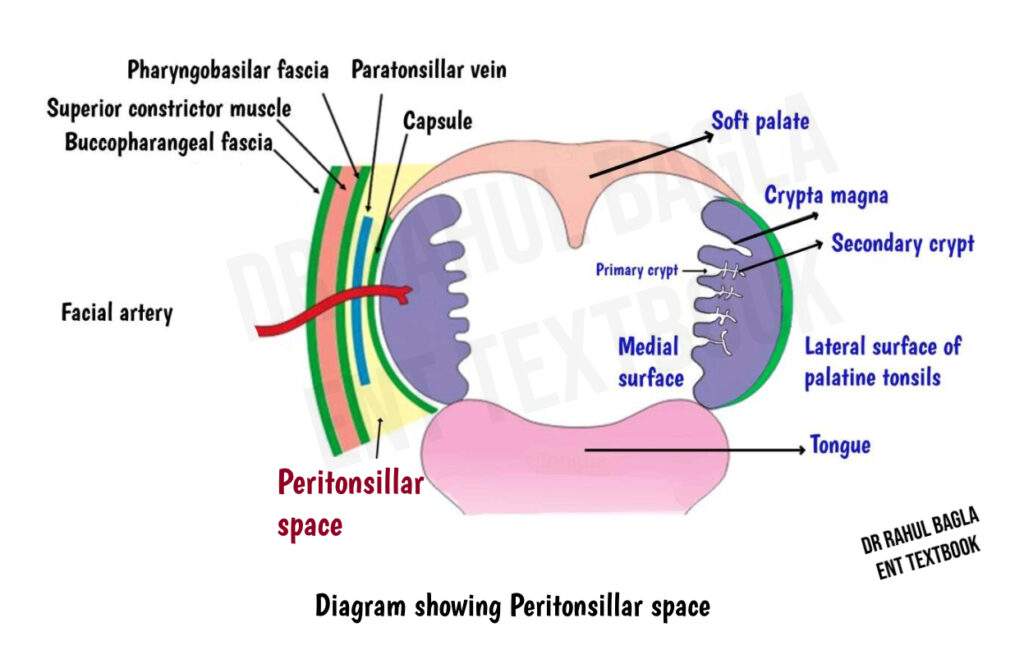
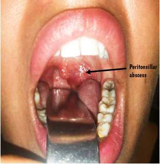
Peritonsillar Abscess: An abscess in this space is called a peritonsillar abscess, commonly known as quinsy. Peritonsillar abscess primarily affects adults, although acute tonsillitis is more common in children. The abscess is usually unilateral, though bilateral cases have been recorded. The infection is usually polymicrobial, involving both aerobic and anaerobic bacteria. Common organisms include Streptococcus pyogenes (the most common aerobic isolate), Fusobacterium necrophorum (an emerging pathogen, found in up to 22.9% of cases), Staphylococcus aureus, and anaerobes such as Prevotella and Peptostreptococcus species.
Aetiology: A peritonsillar abscess usually develops as a complication of acute tonsillitis. Infection commonly begins in a tonsillar crypt, particularly the superior tonsillar crypt (crypta magna) , or from the minor salivary glands of Weber, and may spread through the tonsillar capsule into the peritonsillar space. The infection can also develop independently of acute tonsillitis. If left untreated, it may spread into the parapharyngeal space.
Clinical Features: Patients commonly present with fever, severe unilateral throat pain and odynophagia (pain in swallowing). Severe pain may prevent swallowing of saliva and cause drooling. Other common features include:
- Muffled “hot potato” voice
- Foul-smelling breath
- Trismus due to irritation of the pterygoid muscles
- Tender and enlarged jugulodigastric lymph nodes
- Ipsilateral referred ear pain due to involvement of the glossopharyngeal nerve (CN IX)
Examination: Examination typically shows swelling and congestion of the tonsil, soft palate and tonsillar pillars on the affected side. The soft palate usually bulges above and lateral to the tonsil, and the uvula becomes oedematous and is displaced to the opposite side. Mucopus may be visible over the tonsillar region. Cervical lymphadenopathy commonly involves the jugulodigastric lymph nodes. The patient may hold the head tilted toward the affected side (torticollis) because of pain and muscle spasm.
Differential Diagnosis: Parapharyngeal abscess, Odontogenic abscess (especially upper molar).
Treatment:
- Antibiotics: Early cases of peritonsillar abscess may actually represent peritonsillar cellulitis. Start antibiotics that cover Group A Streptococcus and oral anaerobes, such as penicillin, ampicillin-sulbactam, or clindamycin, usually for about 7 days. Add MRSA coverage with vancomycin or linezolid when MRSA is suspected, or the infection is severe.
- Corticosteroids: A single dose of IV dexamethasone may reduce pain, fever, trismus and hospital stay.
- Drainage: Drainage can be performed by needle aspiration or incision and drainage (I&D). Needle aspiration is both diagnostic and therapeutic, while I&D is commonly used for a first-time abscess. Both are endorsed by AAO-HNS. The intraoral approach is preferred because it provides good access with low morbidity. The incision is made over the most prominent part of the swelling or at the point where a vertical line along the anterior pillar meets a horizontal line drawn at the level of the base of the uvula.
- Supportive care: Give IV fluids and analgesics and advise warm saline mouthwashes.
- Tonsillectomy (AAO-HNS 2019 Guideline): Interval tonsillectomy may be considered in patients with >1 peritonsillar abscess or repeated failed drainage, and is usually performed 4–6 weeks after the acute infection has resolved. Immediate (hot) tonsillectomy may be considered in selected patients with recurrent, bilateral, or difficult-to-drain abscesses.
Complications: An untreated peritonsillar abscess can spread into the parapharyngeal and retropharyngeal spaces. Other complications include airway obstruction, aspiration of pus causing pneumonitis or lung abscess, sepsis, Lemierre syndrome (septic thrombophlebitis of the internal jugular vein with septic emboli), and, rarely, carotid artery or jugular vein erosion causing severe haemorrhage. Intracranial complications such as brain abscess can also occur.
2. Parapharyngeal Abscess. This abscess occurs when the infection spreads from the peritonsillar area into the deeper neck spaces. It poses a risk of descending infection leading to serious complications like mediastinitis.
3. Intratonsillar Abscess. An intratonsillar abscess forms within the tonsil itself, often due to blockage of the crypt opening in cases of acute follicular tonsillitis. It is characterized by marked local pain, swelling, redness, and difficulty in swallowing. Treatment: Administration of antibiotics and drainage of the abscess. Tonsillectomy may be required to prevent recurrence.
4. Tonsilloliths (Tonsil Stones). Tonsilloliths are calcified deposits that form within the tonsillar crypts, commonly in adults. They occur when debris gets trapped in the crypts and calcifies over time, often leading to local discomfort, a foreign body sensation, halitosis, and sore throat.
Diagnosis: They can be identified through palpation or probing, often feeling gritty.
Treatment:
- Conservative treatment includes expressing the tonsilloliths or chemical cauterization of the crypts.
- Tonsillectomy is indicated if there is persistent pain, halitosis, or if the stone is deeply set and cannot be removed easily.
5. Tonsillar cyst. Tonsillar cysts are caused by the blockage of a tonsillar crypt, resulting in a yellowish swelling. They are often asymptomatic but can be easily drained if necessary.
6. Focus of Infection in Systemic Diseases. Chronic tonsillitis can serve as a focal point of infection for systemic diseases such as rheumatic fever, acute glomerulonephritis, and various eye and skin disorders. The persistent infection in the tonsils can trigger these systemic responses, leading to significant health issues beyond the oropharyngeal region.
7. Risks of Recurrent Abscesses. If chronic tonsillitis is not adequately treated, there is a high risk of recurrent abscess formation within the paratonsillar scar tissue, necessitating further medical intervention.
———— End of the chapter ————
Download full PDF Link:
Tonsillitis Best Lecture Notes Dr Rahul Bagla ENT Textbook
Reference Textbooks.
- Scott-Brown, Textbook of Otorhinolaryngology-Head and Neck Surgery.
- Cummings, Otolaryngology-Head and Neck Surgery.
- Stell and Maran’s, Textbook of Head and Neck Surgery and Oncology.
- Ballenger’s, Otorhinolaryngology Head And Neck Surgery
- Susan Standring, Gray’s Anatomy.
- Frank H. Netter, Atlas of Human Anatomy.
- B.D. Chaurasiya, Human Anatomy.
- P L Dhingra, Textbook of Diseases of Ear, Nose and Throat.
- Hazarika P, Textbook of Ear Nose Throat And Head Neck Surgery Clinical Practical.
- Mohan Bansal, Textbook of Diseases of Ear, Nose and Throat Head and Neck Surgery.
- Hans Behrbohm, Textbook of Ear, Nose, and Throat Diseases With Head and Neck Surgery.
- Logan Turner, Textbook of Diseases of The Nose, Throat and Ear Head And Neck Surgery.
- Arnold, U. Ganzer, Textbook of Otorhinolaryngology, Head and Neck Surgery.
- Ganong’s Review of Medical Physiology.
- Guyton & Hall Textbook of Medical Physiology.
Author:

Dr. Rahul Bagla
MBBS (MAMC, Delhi) MS ENT (UCMS, Delhi)
Fellow Rhinoplasty & Facial Plastic Surgery.
Renowned Teaching Faculty
Mail: msrahulbagla@gmail.com
India
———– Follow us on social media ————
- Follow our Facebook page: https://www.facebook.com/Dr.Rahul.Bagla.UCMS
- Follow our Instagram page: https://www.instagram.com/dr.rahulbagla/
- Subscribe to our Youtube channel: https://www.youtube.com/@Drrahulbagla
- Please read. Juvenile Angiofibroma. https://www.entlecture.com/juvenile-angiofibroma/
- Please read. Tumours of Hypopharynx . https://www.entlecture.com/tumours-of-the-hypopharynx/
- Please read. Anatomy of Oesophagus. https://www.entlecture.com/anatomy-of-oesophagus/
Keywords: Acute Follicular tonsillitis, Membranous Tonsillitis, Diphtheria, Vincent’s Angina, Infectious Mononucleosis, Quinsy, Intratonsillar Abscess, Tonsilloliths, Tonsils stones, Tonsillitis symptoms and treatment, how to treat tonsillitis at home, Causes of recurring tonsillitis, Bacterial vs viral tonsillitis differences, When to consider tonsillectomy for tonsillitis, Chronic tonsillitis management tips, Tonsillitis in children: signs and remedies, Natural remedies for tonsillitis relief, Antibiotics for tonsillitis: when are they needed, Complications of untreated tonsillitis, Tonsillitis: Symptoms, Causes, and Effective Treatments, Home Remedies for Tonsillitis: Relieve Pain and Discomfort Naturally, Understanding the Differences Between Bacterial and Viral Tonsillitis, When Does Tonsillitis Require a Tonsillectomy? A Doctor’s Guide, Recurring Tonsillitis: Causes, Prevention, and Treatment Options, Tonsillitis in Children: What Parents Need to Know, Chronic Tonsillitis: Long-Term Management and Care Tips, Antibiotics for Tonsillitis: Do You Really Need Them?, Complications of Tonsillitis: Why Early Treatment Matters, How to Diagnose and Treat Tonsillitis Effectively at Home..