
|
The following CBME core competencies are covered in this chapter.
|
Granulomatous Conditions of the Nose and Paranasal Sinuses
Introduction to Nasal Granulomas
A granuloma represents an organised collection of activated macrophages that forms in response to persistent chronic inflammation. These specialised macrophages transform into epithelioid cells, and many of them fuse together to create multinucleated giant cells, forming the characteristic granulomatous architecture. Granulomas develop in a wide spectrum of infective, inflammatory, and neoplastic conditions affecting the nose and paranasal sinuses, and they typically arise from bacterial infections, fungal infections, or idiopathic causes. Since many granulomatous lesions are intimately associated with systemic diseases, you must always evaluate the patient for systemic involvement at the time of diagnosis.
Clinical Pearl: Any patient with persistent nasal crusting, blood-stained discharge, ulceration, or an atypical nasal mass should raise suspicion for granulomatous disease, chronic infection, or neoplasia and warrants appropriate evaluation.
Biopsy remains essential in all suspected cases because it serves two critical purposes. First, it confirms the diagnosis of granulomatous disease definitively. Second, it helps exclude malignancy, as many of these conditions clinically mimic tumours with alarming similarity. These conditions often present with overlapping clinical features, which makes diagnosis challenging, but their treatment differs significantly. Therefore, accurate identification becomes critical for patient outcomes.
Classification of Granulomatous Diseases
Granulomatous diseases of the nose and paranasal sinuses fall into three major categories based on their underlying aetiology. Understanding this classification helps you approach diagnosis systematically.
| Infective | Immune-mediated / Inflammatory | Neoplastic / Histiocytic | Other Granulomatous Reactions |
| Rhinoscleroma | Granulomatosis with polyangiitis (GPA) | Extranodal NK/T-cell lymphoma (ENKTCL) | Cholesterol granuloma |
| Nasal tuberculosis | Eosinophilic granulomatosis with polyangiitis (EGPA) | Other lymphomas | Foreign-body granuloma |
| Syphilis | Sarcoidosis | Langerhans cell histiocytosis | |
| Leprosy | Cocaine-induced midline destructive lesion (CIMDL) | ||
| Rhinosporidiosis | |||
| Aspergillosis | |||
| Mucormycosis | |||
| Histoplasmosis | |||
| Blastomycosis | |||
| Candidiasis |
Infective Granulomas
Rhinoscleroma
Rhinoscleroma is a chronic, progressive granulomatous condition that primarily affects the nose but can also involve the nasopharynx, larynx, trachea, and bronchi. Laryngeal involvement is called scleroma of the larynx. The disease shows a slight female preponderance and typically affects individuals between 10 and 30 years of age. Rhinoscleroma is a tropical disease with the highest prevalence in Africa and Central America, though cases occur worldwide due to migration. In India, it is seen more often in the northern than in the southern parts.
Aetiology
Klebsiella rhinoscleromatis causes rhinoscleroma. This Gram-negative bacillus belongs to the Enterobacteriaceae family. Anton von Frisch first identified the organism in 1882, hence the alternative name “Frisch bacillus.”
Mode of spread
The bacterium spreads through droplet infection or by inhalation of contaminated material. Once inhaled, it establishes chronic infection in the nasal mucosa, leading to the characteristic granulomatous response.
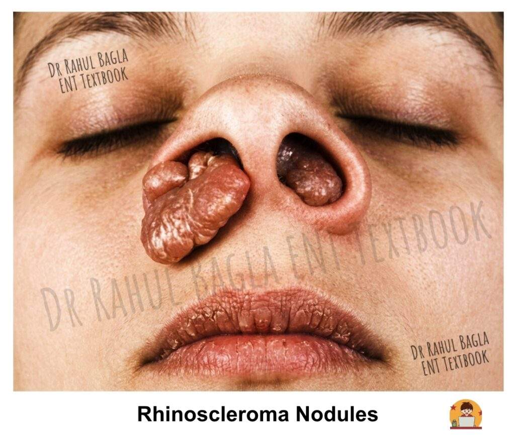
Clinical Stages
The disease progresses through three distinct stages:
- Stage 1: Catarrhal or Atrophic Stage. The catarrhal stage begins with symptoms identical to nonspecific rhinitis. Patients initially experience nasal discharge and obstruction that resemble common cold symptoms. However, as the disease progresses, the nasal discharge becomes purulent and foul-smelling, and crusting develops within the nasal cavity. This stage can easily be mistaken for simple chronic rhinitis or atrophic rhinitis, which often leads to diagnostic delay.
- Stage 2: Granulomatous, Proliferative, or Nodular Stage. During this stage, intranasal rubbery nodules or polyps develop. These lesions are firm on palpation and may bleed easily, causing epistaxis. As the disease advances, destruction of the nasal cartilage occurs, leading to nasal deformity. The external nose becomes swollen and develops a characteristic woody feel because of subdermal infiltration, and this appearance is known as Hebra nose. Furthermore, the external nose becomes enlarged and elongated, resembling the nose of a tapir, hence the term Tapir nose. Laryngeal involvement causes dysphonia; tracheal involvement causes airway obstruction.
- Stage 3: Sclerotic, Cicatricial or Fibrotic Stage. This represents the end stage of the disease, characterised by extensive cicatrisation (the process of scar formation and tissue repair during healing) and fibrosis. The nasal passages become narrowed or stenosed due to the contracture of scar tissue. Patients may have persistent nasal obstruction and cosmetic deformity at this stage, and subglottic stenosis with respiratory distress may also occur.
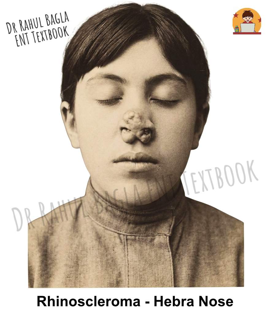
Diagnosis
The diagnosis of rhinoscleroma relies on histopathological examination. Histopathological examination is important for diagnosis. The characteristic findings, particularly in the granulomatous stage, include Mikulicz cells and Russell bodies. Mikulicz cells are large foamy macrophages containing Klebsiella rhinoscleromatis. Russell bodies are eosinophilic immunoglobulin-containing inclusions within plasma cells. In later fibrotic stages, these characteristic cells may be less prominent.
Treatment
- Medical Management. Treatment requires prolonged antimicrobial therapy directed against Klebsiella rhinoscleromatis. Agents such as ciprofloxacin, doxycycline/tetracyclines, rifampicin, or trimethoprim–sulfamethoxazole have been used, with the choice guided by clinical response and antimicrobial susceptibility where available. Treatment is usually continued for an extended period because relapse can occur. Steroids can be combined to reduce fibrosis.
- Surgical Management. Surgical intervention becomes necessary for complications. Dilatation of the nasal cavity may be required for stenosis, and corrective rhinoplasty can address external deformities after the disease is controlled medically.
Key Exam Point: Rhinoscleroma is a classic cause of “Tapir nose” and “Hebra nose.” Mikulicz cells are pathognomonic. Treatment is prolonged antibiotics until two negative cultures.
Nasal Tuberculosis
Aetiology
Mycobacterium tuberculosis, an acid-fast bacillus, is the cause of nasal tuberculosis.
Mode of spread
The infection reaches the nose through three possible routes:
- Direct inoculation by fingernail trauma (a common habit of picking the nose)
- Spread from open pulmonary tuberculosis via infected sputum
- Hematogenous dissemination from a primary infection elsewhere in the body
Clinical Forms and Features
Nasal tuberculosis presents in three main clinical patterns:
- Lupus Vulgaris (Nodular Form). Lupus vulgaris is a low-grade tuberculous infection that typically begins in the nasal vestibule and spreads to adjacent skin and the skin of the nose and face. It results from direct bacterial inoculation into the nasal skin of an immunocompetent patient. Clinically, apple-jelly nodules appear as shiny, reddish-brown papules or nodules. On diascopy, the lesions show the characteristic apple-jelly appearance because pressure causes blanching. The nodules may gradually coalesce and form ulcers with a pale, granular base and undermined edges.
- Ulcerative Form. Nasal TB commonly involves the anterior cartilaginous septum and may lead to septal perforation. Saddle-nose deformity is uncommon, although extensive destructive disease may involve the bony septum. Patients commonly present with nasal obstruction, crusting, discharge, and epistaxis. In contrast, tertiary syphilis characteristically causes destructive involvement of the nasal septum and may produce saddle-nose deformity.
- Sinus Granuloma.Tubercular sinus disease typically presents with diffuse soft-tissue swelling and multiple discharging sinuses. This presentation is usually secondary to bone destruction from osteomyelitis of the frontal bone, and rarely the maxilla or zygomatic bone. CT imaging reveals a soft tissue mass with or without bone destruction.
Diagnosis (Biopsy and Histopathology)
The diagnosis is confirmed by biopsy. Histopathology shows characteristic epithelioid cell granulomas with Langhans-type giant cells, central caseation necrosis, and acid-fast bacilli on Ziehl-Neelsen staining. Microbiological tests include culture on Löwenstein–Jensen medium, which typically takes around 6 weeks. CBNAAT (Cartridge-Based Nucleic Acid Amplification Test) can be performed on pus or discharge from sinuses or nodules, providing rapid diagnosis within hours. The Mantoux test involves purified protein derivative injected intradermally, producing a wheal and flare reaction due to delayed hypersensitivity, and you read the results after 48–72 hours.
When microbiological confirmation is difficult, diagnosis should be based on the overall clinical, histopathological, microbiological, and radiological findings, with specialist consultation where necessary. You should always exclude associated pulmonary tuberculosis by chest X-ray.
Treatment
Nasal tuberculosis responds well to standard antitubercular treatment (ATT). The intensive phase consists of isoniazid, rifampicin, ethambutol, and pyrazinamide for 2 months. The continuation phase involves isoniazid and rifampicin for 4 months. For multidrug-resistant tuberculosis (MDR-TB), treatment is guided by culture-sensitivity tests and requires a longer duration with second-line drugs.
Key Exam Point: Nasal TB spares the bony septum, so it does NOT cause saddle nose deformity. This distinguishes it from syphilis.
Syphilis
Aetiology
Treponema pallidum, a spirochaete, is the cause of syphilis. It can involve the nose at any age. Syphilis remains an important diagnosis and is re-emerging in many parts of the world. Early diagnosis is difficult, especially if patients have received antibiotics for other conditions.
Classification
Syphilis is congenital or acquired. Congenital syphilis presents as early (first 2 years) or late forms; acquired syphilis progresses through primary, secondary, and tertiary stages.
Nasal Manifestations by Stage
- Primary Syphilis. The typical lesion is a chancre appearing 3–4 weeks after initial contact. Chancres are hard, non-tender ulcerated papules that disappear after 6–10 weeks of infection. Common sites are the external nose or vestibule.
- Secondary Syphilis. This stage starts 6–10 weeks after inoculation. Nasal involvement is rare during this stage, though mucous patches may occur.
- Tertiary Syphilis. The characteristic lesion of tertiary syphilis is the gumma, which can involve the mucous membrane, periosteum, and bone. Gummas consist of nodules containing plasma cells, lymphocytes, epithelioid cells, and fibroblasts. Perivascular cuffing and endarteritis by these cells lead to narrowing of the vessel lumen, ultimately causing necrosis and ulceration.
Clinical Features
Patients commonly present with nasal obstruction, crusting, bleeding, and foul-smelling discharge. The disease destroys the bony part of the nasal septum, which leads to the classical saddle nose deformity. This feature clearly distinguishes it from tuberculosis, which spares the bony septum. Patients may also report nasal tenderness and headache.
Complications
Advanced disease can cause septal perforation, palatal and nasal wall perforation, sequestrum formation, and collapse of the nasal bridge. Progressive scarring may lead to nasal stenosis and atrophic rhinitis. In rare cases, infection may spread intracranially.
Diagnosis
It requires a combination of nontreponemal tests (RPR or VDRL) and treponemal tests (TPPA, FTA-ABS, EIA/CIA, or equivalent). Depending on the laboratory algorithm, either a nontreponemal or treponemal test may be used initially.
Treatment
- Primary, secondary, and early latent syphilis: Benzathine penicillin G 2.4 million units IM as a single dose.
- Late latent syphilis or syphilis of unknown duration: Benzathine penicillin G 2.4 million units IM weekly for 3 doses (total 7.2 million units).
- Tertiary syphilis: Requires assessment for cardiovascular, gummatous, and neurological involvement; treatment depends on the clinical form.
- Neurosyphilis, ocular syphilis, and otosyphilis: Require an appropriate aqueous crystalline penicillin G regimen, generally administered intravenously.
- Penicillin allergy: Doxycycline 100 mg BD for 14 days (early) or 28 days (late latent/tertiary). but penicillin remains the preferred.
- Local nasal care: Gentle cleansing and saline irrigation may help remove crusts and improve symptoms.
Sequelae Management. Atrophic rhinitis and deformity may persist after the disease and require appropriate medical and surgical management.
Key Exam Point: Syphilis destroys the bony septum, causing saddle nose deformity. VDRL is the screening test; FTA-ABS and TPHA are confirmatory.
Leprosy (Hansen’s Disease)
Aetiology
Leprosy (Hansen’s disease) is a chronic infection caused mainly by Mycobacterium leprae, an acid-fast bacillus. It has a long incubation period of 3-10 years.
Classification
Leprosy forms a spectrum based on host immunity (Ridley-Jopling classification):
- Tuberculoid (high immunity) – At the tuberculoid end, patients show few skin lesions, and microscopy reveals absent or very few bacilli. This form reflects strong immunity, so it remains the least contagious and relatively benign.
- Borderline tuberculoid
- Mid-Borderline
- Borderline lepromatous
- Lepromatous (low immunity) – At the lepromatous end, patients develop multiple widespread skin lesions filled with numerous bacilli. This pattern indicates poor immunity, making the disease more severe and highly infectious.
Unique Features of M. leprae
Leprae is unique among bacteria because it selectively invades peripheral nerves, causing irreversible cutaneous anaesthesia and paralysis of muscles in the extremities and face. The earlobes and nose are especially enlarged and infiltrated in lepromatous leprosy. Widespread facial skin lesions give rise to the characteristic Leonine facies (lion-like face).
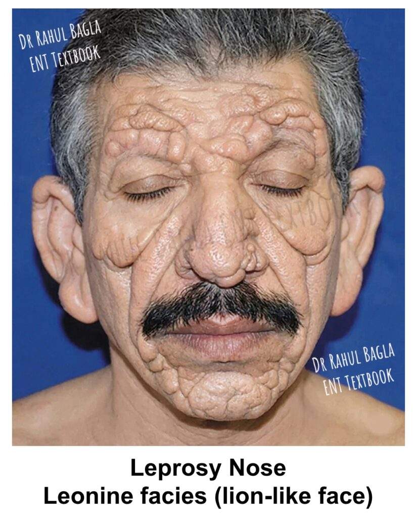
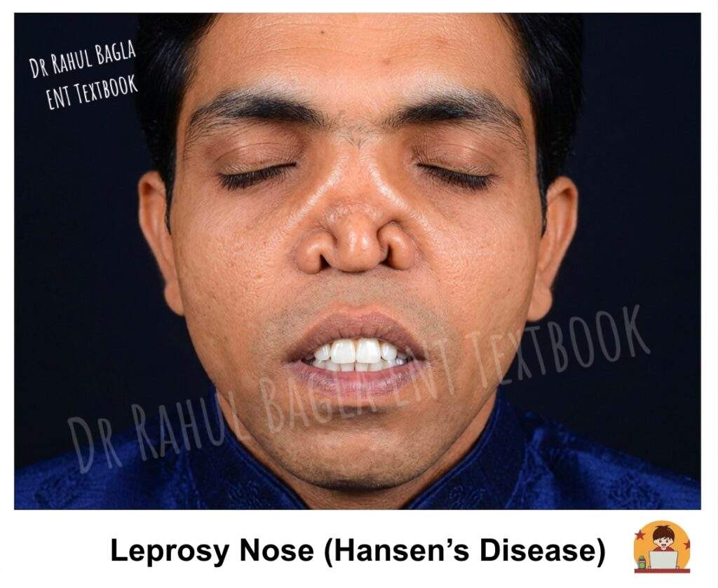
Clinical Features
Lepromatous leprosy involves the nose early in the disease course. Patients commonly present with nasal obstruction, crusting, bleeding, and a reduced sense of smell (hyposmia). The disease may also affect other head and neck sites such as the auricle, hard and soft palate, epiglottis, and larynx. Laryngeal leprosy can cause airway obstruction and requires urgent attention. On examination, the nasal mucosa shows pale yellowish thickening with nodules or plaques. As the disease progresses, it produces diffuse nodular infiltration, especially over the inferior turbinate and nasal septum. In advanced stages, patients may develop septal perforation, saddle nose deformity, and atrophic rhinitis.
Treatment
Treatment is long-term multidrug therapy (MDT). According to current WHO recommendations, the regimen consists of rifampicin, dapsone, and clofazimine, with a treatment duration of 6 months for paucibacillary (PB) leprosy and 12 months for multibacillary (MB) leprosy. Alternative agents: Minocycline, clarithromycin, and quinolones such as ofloxacin may be used in selected cases, particularly when standard MDT cannot be used or in drug-resistant leprosy.
Key Exam Point: Lepromatous leprosy causes Leonine facies, saddle nose deformity, and nasal crusting. MDT duration: 6 months PB, 12 months MB.
Rhinosporidiosis
Epidemiology
Rhinosporidiosis is a chronic mucosal infection that is endemic in parts of South Asia, particularly India and Sri Lanka, with cases also reported from other tropical and subtropical regions. It is a classic exam topic due to its geographic predilection.
Aetiology
Rhinosporidiosis is caused by Rhinosporidium seeberi, historically classified as a fungus. Molecular studies have placed it within the Mesomycetozoea, a group of aquatic protists, rather than among true fungi.
Mode of spread
This organism is associated with stagnant or slow-moving freshwater, and infection is thought to occur through transepithelial penetration of the organism into mucosal surfaces, particularly after exposure to contaminated water. Minor mucosal trauma or abrasions may facilitate entry, although the exact mode of transmission remains uncertain. The disease also occurs in animals, including cattle, horses, and dogs, supporting an environmental reservoir or common source of exposure.
Pathogenesis and Life Cycle
The organism undergoes progressive development within the host tissue, culminating in the formation and rupture of mature sporangia.
- Endospore and Trophocyte Stage: The cycle begins when a small endospore, approximately 5–10 μm in diameter, enters the mucosa or submucosal tissue. This endospore, with a distinct chitinous wall, contains a clear cytoplasm and a vesicular nucleus. It then begins to grow, undergoing multiple nuclear and cytoplasmic divisions. The trophocyte enlarges, filled with young endospores.
- Sporangium Development: The developing trophocyte enlarges and forms a large, thick-walled sporangium. These sporangia are the defining histopathological feature of the disease, measuring a massive 200–250 μm in diameter – a size that can be seen with a low-power microscope. Each sporangium is filled with thousands (up to 16,000) of endospores.
- Endospore Production and Release: The mature sporangium eventually ruptures, releasing infective endospores into the surrounding tissue. These liberated endospores then begin the new cycle, leading to local spread and recurrence. Rarely, endospores disseminate via lymphatics or blood.
Clinical Features
Rhinosporidiosis can occur at any age but is particularly common among children, adolescents, and young adults, with a male predominance (4:1) reported in many series. The disease is characterised by a persistent, slow-growing, polypoidal mass in the nasal cavity, though it can affect other sites.
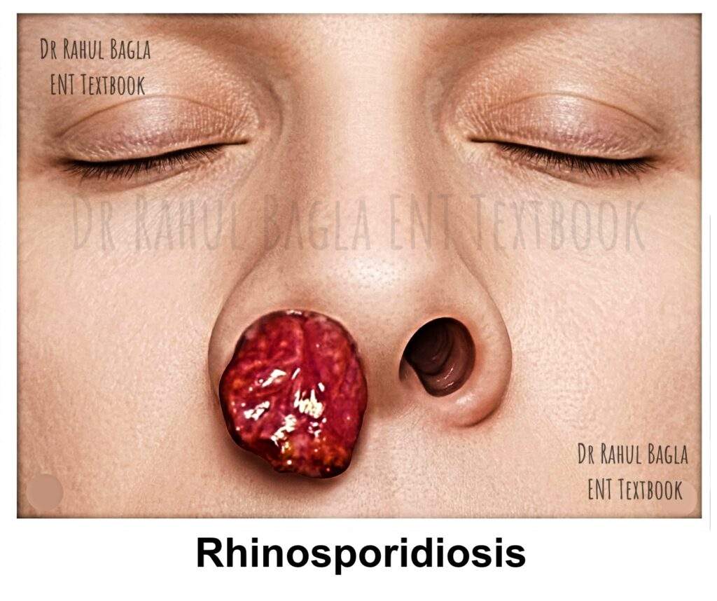
- Nasal Manifestations: The nose is the most common site of involvement. The classic presentation is a fleshy, strawberry-like mass. This vascular, friable polyp is pink to purple and attached to the nasal septum or lateral wall. Its surface is studded with characteristic yellowish-white or greyish-white dots, representing mature sporangia visible through the overlying epithelium. The mass is highly vascular and friable and tends to bleed easily on manipulation. Patients have progressive nasal obstruction and blood-stained discharge.
- Extranasal Manifestations: While the nose is the primary site, the disease can occasionally involve the nasopharynx, lips, palate, uvula, conjunctiva, larynx, and even the genitals. Therefore, a diagnosis may be delayed when the disease presents at these less common sites.
Diagnosis and Histopathology
Diagnosis is primarily clinical, supported by histopathological examination of the excised mass. Biopsy is essential but must be done with caution due to the vascular nature of the lesion.
- Gross Appearance: A fleshy, pedunculated or sessile polyp with a characteristic strawberry-like surface.
- Microscopy: H&E shows pathognomonic sporangia in various stages. These large, thick-walled structures containing endospores are diagnostic. Fungal stains like Gomori methenamine silver (GMS) and periodic acid-Schiff (PAS) can also highlight the organism. A chronic inflammatory reaction with epithelioid cell granulomas and giant cells is seen in about 50% of cases.
Treatment
Treatment is primarily surgical, as medical therapy has a limited role.
- Surgical Excision: Complete excision of the mass with electrocoagulation of its base is the treatment of choice to prevent recurrence. Endoscopic endonasal surgery is the preferred approach, as it allows for precise, complete excision and good visualisation of the base.
- Medical Therapy: Dapsone has been used as an adjunct to surgery because it appears to inhibit maturation of sporangia and promote stromal fibrosis. However, evidence for its effectiveness in preventing recurrence is limited and variable. It may be considered in recurrent, extensive, or disseminated disease, under specialist supervision.
Key Exam Point: Rhinosporidiosis is endemic in southern India and Sri Lanka. The strawberry-like mass with greyish-white dots is pathognomonic. Treatment is complete surgical excision with electrocoagulation. Biopsy is NOT contraindicated but must be done with caution.
Aspergillosis
Aspergillosis is caused by several species of Aspergillus, most commonly A. fumigatus and A. flavus. The clinical presentation depends on the host’s immune status, underlying disease, and the type of sinonasal involvement.
Sinonasal aspergillosis can present in four major forms:
- Fungus Ball (Non-invasive): This is a common form of sinonasal aspergillosis and typically occurs in immunocompetent individuals. It usually involves a single sinus, most commonly the maxillary sinus, and often presents as chronic unilateral sinonasal disease. The sinus contains a dense collection of fungal hyphae, mucus, cellular debris, and mineral deposits forming a firm fungal ball (mycetoma). On endoscopy, the material may appear dark brown, greenish, or black. Treatment is surgical removal alone; antifungal medication is not routinely required for an uncomplicated fungus ball.
- Allergic Fungal Rhinosinusitis (AFRS): Occurs predominantly in immunocompetent, atopic individuals and is characterised by chronic rhinosinusitis, nasal polyps, and thick eosinophilic mucin containing fungal hyphae. CT typically shows heterogeneously hyperdense sinus contents, sometimes producing the characteristic “double-density” appearance, along with sinus expansion and bony remodelling or erosion in some cases. Treatment is primarily endoscopic removal of polyps and allergic mucin, followed by topical intranasal corticosteroids; systemic corticosteroids may be used selectively in severe disease.
- Chronic Invasive Aspergillosis: This is a slowly progressive form of invasive fungal sinusitis that may occur in patients with mild or moderate immunosuppression or certain underlying diseases. It can produce progressive destruction of the sinonasal tissues and may extend into the orbit or adjacent structures.
- Acute Invasive (Fulminant) Aspergillosis: Occurs predominantly in severely immunocompromised patients, particularly those with prolonged neutropenia, haematological malignancy, transplantation, or significant corticosteroid exposure. It is an aggressive angioinvasive infection that can rapidly cause tissue necrosis and destruction. Clinical features may include fever, facial pain or swelling, nasal obstruction, ophthalmic symptoms, and necrotic or discoloured nasal mucosa. Treatment requires urgent surgical debridement when feasible, together with systemic antifungal therapy. Voriconazole is a preferred first-line agent for invasive aspergillosis, while liposomal amphotericin B or isavuconazole are important alternatives depending on the clinical situation.
Key Exam Point: Sinonasal aspergillosis may present as a fungus ball, allergic fungal rhinosinusitis, chronic invasive disease, or acute invasive disease. Fungus ball is treated primarily by surgical removal, AFRS by endoscopic clearance followed by medical control of inflammation, and invasive disease requires systemic antifungal therapy with surgical debridement when indicated.
Mucormycosis
Mucormycosis is a rapidly progressive and potentially fatal fungal infection caused by fungi of the order Mucorales (e.g., Rhizopus, Mucor). It is associated with uncontrolled diabetes mellitus, especially DKA. Other risk factors include haematological malignancies, stem cell transplantation, prolonged corticosteroid use, and deferoxamine therapy.
A hallmark of mucormycosis is angioinvasion. Broad, irregular fungal hyphae invade blood-vessel walls, producing thrombosis, vascular occlusion, tissue ischemia, and extensive necrosis. This may produce a black or dark necrotic eschar in the nasal cavity or on the palate, reflecting tissue ischemia and necrosis. Although highly suggestive in the appropriate clinical setting, a black eschar is not pathognomonic of mucormycosis.
Rhino-orbital disease may extend from the sinonasal tract into the orbit and, in advanced cases, intracranially. Clinical manifestations may include facial pain or swelling, orbital oedema, proptosis, ophthalmoplegia, visual loss, and neurological complications. Palatal involvement may result in tissue necrosis and perforation.
Treatment
Management is a medical and surgical emergency and requires early diagnosis, assessment of disease extent, aggressive surgical debridement, prompt antifungal therapy, and reversal of underlying predisposing factors.
- Assessment of Disease Extent: Urgent CECT PNS and/or MRI to assess sinonasal, orbital, and intracranial involvement. Imaging should not delay treatment when clinical suspicion is high.
- Urgent Surgical Debridement: Prompt surgical debridement of necrotic tissue. Repeated debridement may be required, depending on disease progression and the presence of residual necrotic tissue.
- Antifungal Therapy: Liposomal amphotericin B is the preferred first-line antifungal therapy, generally administered at 5–10 mg/kg/day, with the dose selected according to disease severity and clinical circumstances. Amphotericin B deoxycholate is generally avoided because of its greater nephrotoxicity. Posaconazole and isavuconazole are alternatives for step-down therapy or when Amphotericin B is contraindicated.
- Correction of Predisposing Factors: This is critical for successful treatment.
Inflammatory Granulomas
Granulomatosis with Polyangiitis (GPA)
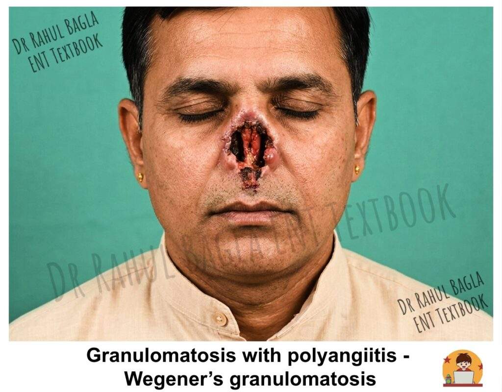
Granulomatosis with polyangiitis (GPA), formerly known as Wegener’s granulomatosis, is a systemic, autoimmune, ANCA-associated necrotising granulomatous vasculitis of small- and medium-sized vessels. It classically involves the upper respiratory tract, lower respiratory tract, and kidneys, but the complete triad is present in only a minority of patients.
Epidemiology
Uncommon. Can occur at any age; most common in middle adulthood. No ethnic predilection.
Aetiology and Pathogenesis
GPA is strongly associated with antineutrophil cytoplasmic antibodies (ANCA), particularly proteinase 3 (PR3)-ANCA, which commonly produces a cytoplasmic (c-ANCA) pattern on indirect immunofluorescence. PR3-ANCA can activate primed neutrophils, contributing to endothelial injury and necrotising inflammation. However, ANCA is not detected in every patient, particularly in patients with disease confined to the upper respiratory tract. Antigen-specific immunoassays for PR3-ANCA and MPO-ANCA are preferred for laboratory evaluation. Nasal carriage of Staphylococcus aureus has been associated with an increased risk of relapse, although the mechanism and clinical implications remain incompletely understood.
Clinical Features
- Nasal/sinus (>80%): Crusting, epistaxis, nasal obstruction, purulent discharge. Progressive destruction may cause septal perforation, saddle-nose deformity, and a single large nasal cavity (“autorhinectomy”).
- Aural: Otitis media with effusion, conductive or sensorineural hearing loss; facial nerve palsy (uncommon).
- Oral: Ulceration; “strawberry gingivitis hyperplasia”, where the gums appear friable and hyperplastic, is characteristic but not pathognomonic.
- Laryngeal: Subglottic or tracheal stenosis – suspect GPA in any adult with apparently idiopathic subglottic stenosis.
- Ocular: Scleritis, episcleritis, uveitis, dacryocystitis; retro-orbital disease can cause proptosis or visual loss.
- Pulmonary: Cough, hemoptysis, cavitating nodules.
- Renal: Rapidly progressive glomerulonephritis (which can lead to renal failure)
- Neurological: Mononeuritis multiplex, cranial neuropathy.
Diagnosis
Diagnosis is based on clinical features, serology, imaging, and histopathology. No single test is definitive.
- Serology: PR3-ANCA is strongly associated with GPA; MPO-ANCA is more typical of MPA. Modern testing uses antigen-specific immunoassays. A negative ANCA does not exclude GPA — sensitivity is lower in localised sinonasal disease. Also check FBC, ESR, CRP, renal function, urinalysis, and chest imaging.
- Histopathology: Necrotising granulomatous inflammation and vasculitis. The complete triad is not always present. Sinonasal biopsies have limited sensitivity.
- Imaging: CT shows mucosal thickening, bone destruction, sclerosis, and neo-osteogenesis. MRI is useful for orbital and intracranial assessment. Findings support but do not confirm the diagnosis.
Treatment
Treatment is directed at achieving remission, preventing irreversible organ damage, and maintaining remission while minimising treatment-related toxicity. Management should generally involve rheumatology and, when appropriate, nephrology, pulmonology, ophthalmology, and ENT specialists.
- Remission Induction. For organ- or life-threatening GPA, induction therapy generally consists of glucocorticoids plus rituximab or cyclophosphamide. Avacopan, a C5a receptor inhibitor, may be used as a glucocorticoid-sparing strategy in selected patients. For non-organ-threatening disease, methotrexate with glucocorticoids is an established induction option. Other immunosuppressive agents may be considered when standard therapy is unsuitable.
- Remission Maintenance. Rituximab is generally preferred for maintenance of remission. Azathioprine or methotrexate are alternatives in appropriate patients. Maintenance therapy is usually continued for several years, with duration individualised according to relapse risk, disease phenotype, treatment response, and adverse effects.
- Monitoring. Disease activity should be monitored using clinical assessment and organ-specific investigations, including renal function and urinalysis when appropriate. ANCA titres may provide supportive information but should not be used alone to determine disease activity or trigger treatment changes.
- Symptomatic Management: This includes nasal saline douching, topical steroids, and endoscopic sinus surgery to manage sinus disease. Surgical repair of a saddle-nose deformity or septal perforation is only performed when the disease has been in remission for at least one year.
Eosinophilic Granulomatosis with Polyangiitis (EGPA)
EGPA (formerly Churg-Strauss syndrome) is a vasculitis of small and medium vessels. It causes eosinophil-rich inflammation and necrotising granulomas. It is strongly linked with asthma and high eosinophil counts. ANCA is not positive in all patients — many cases are ANCA-negative.
Epidemiology
Rare. Most often diagnosed in middle age. Can occur at any age. No clear sex difference.
Pathogenesis
The exact cause is unknown. It is likely an autoimmune disease where Th2 cells activate eosinophils and neutrophils. Eosinophilic infiltration and release of cationic proteins damage tissues. Zafirlukast (a leukotriene antagonist) has been linked with EGPA. However, this is now thought to be due to unmasking of existing disease when steroids are reduced, not a direct cause.
Clinical Features
Asthma occurs in most patients and is usually adult-onset. It typically comes years before the vasculitis. ENT involvement is very common — allergic rhinitis and CRS with or without polyps. Patients have nasal obstruction, rhinorrhoea, anosmia, sneezing, crusting, and epistaxis. Pulmonary infiltrates are often transient and migratory. Neurological: mononeuritis multiplex, peripheral neuropathy. Skin: papules and nodules. Cardiac involvement (granulomatous myocarditis) is the main cause of death.
Three phases:
- Prodromal — asthma, rhinitis, CRS
- Eosinophilic — tissue and blood eosinophilia, lung infiltrates
- Vasculitic — systemic vasculitis with fever, myocarditis, neuropathy, skin lesions
Diagnosis
Histology shows necrotising vasculitis, extravascular necrotising granulomas, and tissue eosinophilia – but rarely all three in one biopsy. Skin, nerve, and muscle are preferred biopsy sites. ANCA is positive in only some patients, usually MPO-ANCA. ANCA-positive disease tends to have more renal and neurological involvement. ANCA-negative disease is more often linked with cardiac and eosinophilic features. CXR may show transient peripheral infiltrates.
Treatment.
- Non-organ-threatening disease: Systemic glucocorticoids are the mainstay of initial treatment. Mepolizumab (anti-IL-5) is an important steroid-sparing option, particularly in relapsing or refractory disease. Methotrexate, azathioprine, or mycophenolate mofetil may be considered in selected patients.
- Organ- or life-threatening disease: Systemic glucocorticoids combined with cyclophosphamide or rituximab are used for remission induction.
- Relapsing or refractory non-organ-threatening disease: Mepolizumab is an established treatment option and may reduce glucocorticoid exposure.
ENT Pearl: Adult-onset asthma + nasal polyps + high eosinophils should raise suspicion for EGPA. Sinonasal symptoms are often the first sign.
Cocaine-Induced Midline Destructive Lesion (CIMDL)
CIMDL is a destructive midline lesion caused by intranasal cocaine use, often due to levamisole adulteration. It can mimic localised GPA or cancer.
Clinical Features
Chronic nasal obstruction, bleeding, change in nasal shape, and nasal regurgitation as the lesion destroys the nasal framework and palate. Palate perforation is more common than in GPA. Neuropathic pain is common. Systemic symptoms are usually absent. Not all patients admit cocaine use.
Diagnosis
Diagnosis requires combining exposure history, clinical pattern, histology, imaging, and toxicology. HNE-ANCA (p-ANCA against human neutrophil elastase) is a characteristic finding but is not definitive; overlap with GPA occurs. Urine, blood, or hair can be tested for cocaine.
Treatment
For isolated CIMDL, the main treatment is complete cessation of cocaine; immunosuppression is generally not needed. Patients with systemic autoimmune features need multidisciplinary assessment and may need immunosuppression. Reconstructive surgery should wait until the patient has stopped cocaine and the disease is stable. The exact timing depends on the individual patient.
Sarcoidosis
Sarcoidosis is a multisystem granulomatous disorder of unknown aetiology, characterised by the formation of non-caseating granulomas. While the lungs and lymph nodes are most commonly involved, sinonasal sarcoidosis occurs in a small percentage of patients (<5%).
Aetiology and Pathology
The exact cause of sarcoidosis is unknown, but it is thought to result from an exaggerated immune response to an unknown antigen in genetically susceptible individuals. The hallmark is the presence of non-caseating epithelioid cell granulomas on histopathology. This is the key feature that distinguishes it from tuberculosis.
Clinical Features
Sinonasal involvement can manifest in various ways:
- Nasal Symptoms: Nasal obstruction, crusting, epistaxis, and facial pain are common.
- Examination: The nasal mucosa may appear pale and hypertrophied. Multiple, small, pale granulomas can give the mucosa a “cobblestone” or granular appearance, sometimes referred to as “strawberry skin.” This can lead to ulceration, crusting, and eventually, septal perforation.
- Cutaneous Manifestations: Lupus pernio is a characteristic cutaneous manifestation of sarcoidosis, presenting as violaceous, indurated plaques on the nose, cheeks, and ears.
- Associated Syndromes: Heerfordt’s syndrome (uveoparotid fever) is a classic presentation involving parotid gland enlargement, facial nerve palsy, uveitis, and fever.
Diagnosis and Staging
Diagnosis is based on a combination of clinical, radiographic, and histopathological findings, with the exclusion of other granulomatous diseases like tuberculosis.
- Histopathology: Biopsy of the nasal lesion shows the pathognomonic non-caseating granulomas.
- Serology: Serum angiotensin-converting enzyme (ACE) levels are elevated in approximately 85% of patients with active disease. Serum and urinary calcium levels may also be elevated.
- Imaging: A chest X-ray or CT scan is crucial, often revealing bilateral hilar lymphadenopathy (BHL) and pulmonary infiltrates. A staging system (Stages I-IV) is used for pulmonary sarcoidosis. For sinonasal involvement, a simple staging system exists: Stage I (reversible nasal disease), Stage II (moderate, potentially reversible disease with sinus involvement), and Stage III (severe, irreversible, extensive disease).
Treatment
The course of sarcoidosis is variable, and many cases are self-limiting.
- Medical Therapy: Corticosteroids are the mainstay of treatment for symptomatic or progressive disease. For chronic or steroid-resistant cases, steroid-sparing agents like methotrexate, azathioprine, or hydroxychloroquine (especially for skin and sinonasal disease) are used.
- Supportive Care: For sinonasal symptoms, regular nasal douching with saline is essential to manage crusting. Glucose and glycerine drops can help moisturise the nasal mucosa. Surgery is reserved for cosmetic correction of nasal deformities (e.g., saddle-nose) only when the disease is inactive.
Neoplastic Granuloma
Extranodal NK/T-Cell Lymphoma (ENKTCL)
Extranodal NK/T-cell lymphoma (ENKTCL), previously known by many names including “lethal midline granuloma” and “polymorphic reticulosis,” is a rare, aggressive type of non-Hodgkin lymphoma. It is characterised by a destructive, necrotic lesion of the midface.
Aetiology and Pathology
ENKTCL is strongly associated with the Epstein-Barr virus (EBV). It arises from natural killer (NK) cells or, less commonly, T-cells. The defining histological features are an angiocentric and angioinvasive growth pattern, where the neoplastic lymphoid cells infiltrate and destroy blood vessel walls, leading to massive tissue necrosis.
Clinical Features
The disease typically presents in middle-aged to older adults (5th-6th decade) with a male predominance.
- Localised Disease: The disease usually starts unilaterally in the nasal cavity. Patients present with progressive nasal obstruction, purulent and often foul-smelling discharge, and severe epistaxis.
- Midfacial Destruction: The hallmark is relentless, progressive destruction of the midface. It can erode through the nasal septum, hard palate, and the skin of the nose and face, creating a large, central facial defect. Unlike GPA, there is no associated renal or pulmonary involvement in the early stages.
- Systemic Involvement: The disease can disseminate to involve the skin, gastrointestinal tract, and other sites.
Diagnosis and Differential Diagnosis
A high index of suspicion is necessary, as the early presentation mimics chronic sinusitis.
- Histopathology and Immunohistochemistry: Multiple, deep biopsies are required to make the diagnosis. The tissue shows a dense, atypical lymphoid infiltrate with angioinvasion and extensive necrosis. Immunohistochemistry is essential and typically shows positivity for CD56 (a marker for NK cells), CD2, and cytoplasmic CD3ε. The tumour cells are also EBV-positive.
- Imaging: CT and MRI are crucial to assess the extent of local destruction and for staging the disease.
- Differential Diagnosis: It is critical to differentiate ENKTCL from GPA, as the treatments are entirely different. The absence of renal, pulmonary, and c-ANCA positivity, coupled with the aggressive local destruction, points towards ENKTCL.
Treatment
The management of ENKTCL has evolved, and it is no longer a uniformly fatal disease.
- Combined Modality Therapy: The current standard of care for localised ENKTCL is combined chemotherapy and radiotherapy. “Sandwich” regimens, where patients receive a few cycles of chemotherapy (e.g., SMILE protocol: dexamethasone, methotrexate, ifosfamide, L-asparaginase, etoposide) followed by radiation, are effective. For disseminated disease, chemotherapy is the mainstay.
Table: Differential Diagnosis of Granulomatous and Destructive Sinonasal Conditions
| Disease | Aetiology | Typical Sinonasal Pattern | Key Clinical / Systemic Clue | Characteristic Histopathology / Diagnostic Clue | Treatment |
| Rhinoscleroma | Klebsiella rhinoscleromatis | Chronic granulomatous → fibrotic disease with progressive obstruction and stenosis | May extend to nasopharynx, larynx and tracheobronchial tree | Mikulicz cells + Russell bodies; bacterial identification may support diagnosis | Ciprofloxacin or doxycycline/tetracycline |
| Nasal tuberculosis | Mycobacterium tuberculosis | Ulcerative or granulomatous lesion, usually involving the anterior nasal septum | May coexist with pulmonary or extrapulmonary TB | Caseating granulomas ± Langhans giant cells; AFB or molecular testing may confirm | Antituberculous therapy (ATT) |
| Syphilis | Treponema pallidum | Destructive, gummatous disease, particularly in tertiary syphilis | Bony destruction, palatal perforation and saddle-nose deformity | Plasma-cell-rich inflammation, obliterative endarteritis and gumma; treponemal serology | Benzathine penicillin G |
| Leprosy | Mycobacterium leprae | Diffuse mucosal infiltration, crusting and progressive tissue destruction | Peripheral neuropathy, sensory loss and characteristic skin lesions | Granulomatous inflammation with variable acid-fast bacilli, depending on disease spectrum | Multidrug therapy (MDT) |
| Rhinosporidiosis | Rhinosporidium seeberi | Friable, highly vascular polypoidal mass | Usually localized mucosal disease; freshwater exposure is typical | Large thick-walled sporangia containing numerous endospores | Surgical excision + electrocoagulation/cauterisation |
| Aspergillosis | Aspergillus spp. | Fungus ball, allergic fungal rhinosinusitis or invasive sinonasal disease | Invasive disease occurs particularly with immunosuppression; AFRS is associated with atopy | Septate hyphae with acute-angle branching; tissue invasion distinguishes invasive disease | Surgery ± corticosteroids; systemic antifungals for invasive disease |
| Mucormycosis | Mucorales, especially Rhizopus spp. | Rapidly progressive necrotising rhinosinusitis | Diabetes, especially DKA, or severe immunosuppression; orbital involvement may occur | Broad, pauciseptate hyphae with irregular/right-angle branching and angioinvasion | Surgical debridement + liposomal amphotericin B |
| Granulomatosis with polyangiitis (GPA) | Autoimmune ANCA-associated vasculitis | Chronic destructive rhinosinusitis with crusting, ulceration and septal perforation | Pulmonary and renal involvement; PR3-ANCA commonly associated | Necrotising granulomatous inflammation ± vasculitis; PR3-ANCA supports diagnosis | Glucocorticoids + rituximab/cyclophosphamide |
| Eosinophilic granulomatosis with polyangiitis (EGPA) | Eosinophil-rich ANCA-associated vasculitis | Chronic rhinosinusitis, often with nasal polyps | Adult-onset asthma + eosinophilia; neuropathy and cardiac disease may occur | Eosinophil-rich inflammation, necrotising granulomas and/or vasculitis; MPO-ANCA in a subset | Glucocorticoids ± mepolizumab |
| Cocaine-induced midline destructive lesion (CIMDL) | Chronic intranasal cocaine use, often involving levamisole-adulterated cocaine | Severe midline destructive lesion with septal ± palatal perforation | History of cocaine exposure; systemic vasculitic features are usually absent | Necrosis and mixed inflammation; HNE-ANCA may support the diagnosis but is not specific | Cocaine cessation |
| Sarcoidosis | Multisystem granulomatous inflammatory disease of uncertain cause | Chronic crusting, obstruction, granular or nodular nasal mucosa | Pulmonary disease, bilateral hilar lymphadenopathy or lupus pernio | Non-necrotising granulomas after exclusion of infection and other granulomatous disorders | Glucocorticoids ± steroid-sparing therapy |
| Extranodal NK/T-cell lymphoma, nasal type (ENKTCL) | EBV-associated NK/T-cell lymphoma | Aggressive necrotising and destructive midline lesion | Rapid progression; may involve skin, gastrointestinal tract or other extranodal sites | Angiocentric/angiodestructive atypical lymphoid infiltrate + extensive necrosis + EBER positivity | Radiotherapy + chemotherapy |
——– End of the Chapter——–
High-Yield Points for Revision
- Rhinoscleroma — Tapir nose, Hebra nose, Mikulicz cells (foamy macrophages with K. rhinoscleromatis), Russell bodies. Treatment: prolonged antibiotics.
- Nasal TB — Anterior cartilaginous septum involved; saddle-nose is uncommon (unlike syphilis). Apple-jelly nodules on diascopy in lupus vulgaris. AFB may be sparse.
- Syphilis — Destroys bony septum → classic saddle-nose deformity. Diagnosis uses both nontreponemal (VDRL/RPR) and treponemal (TPHA/FTA-ABS/TPPA) tests.
- Leprosy — Leonine facies + saddle-nose + thickened earlobes. Nose involved early in lepromatous form. MDT: 6 months PB, 12 months MB.
- Rhinosporidiosis — Strawberry-like friable polyp with yellowish-white dots (sporangia). R. seeberi is a Mesomycetozoan protist, not a fungus. Treatment: surgical excision + cauterisation.
- Aspergillosis — Four forms: fungus ball (surgery alone), AFRS (surgery + topical steroids), chronic invasive, acute invasive (voriconazole + surgery).
- Mucormycosis — DKA + black necrotic eschar + angioinvasion. Broad pauci-septate hyphae. Treatment: urgent debridement + liposomal amphotericin B + correct DKA. Black eschar is NOT pathognomonic.
- GPA — ANCA-associated necrotising granulomatous vasculitis. Triad: upper respiratory + lower respiratory + renal (but triad not always present). PR3-ANCA strongly associated. Treatment: steroids + rituximab or cyclophosphamide; rituximab for maintenance.
- EGPA — Asthma + eosinophilia + vasculitis. p-ANCA (anti-MPO) in a subset. Treatment: steroids ± mepolizumab for relapsing/refractory non-severe disease.
- CIMDL and ENKTCL — CIMDL: cocaine-induced midline destruction; HNE-ANCA supports but is not definitive; treatment is cocaine cessation. ENKTCL: EBV+ (EBER+), CD56+, angiocentric lymphoma; treat with chemo-radiotherapy. Do not use absence of c-ANCA to distinguish from GPA.
NEET PG MCQs
- A 25-year-old man from northern India presents with a woody, swollen external nose resembling a tapir’s nose. Biopsy shows large foamy macrophages containing bacilli. What is the most likely diagnosis? A) Nasal tuberculosis B) Rhinoscleroma C) Leprosy D) Sarcoidosis
- A 30-year-old woman presents with a friable, strawberry-like nasal polyp studded with yellowish-white dots. It bleeds on touch. What is the treatment of choice? A) Systemic amphotericin B B) Complete surgical excision with cauterisation of base C) Tetracycline for 6 weeks D) Radiotherapy
- A 55-year-old diabetic in ketoacidosis has a black necrotic eschar over the palate and right nasal cavity, with proptosis and ophthalmoplegia. What is the most appropriate next step? A) Start oral fluconazole B) Urgent surgical debridement with liposomal amphotericin B C) Prescribe broad-spectrum antibiotics D) Order an MRI brain and await results
- A 45-year-old man has saddle-nose deformity, chronic nasal crusting, haemoptysis, and haematuria. Which investigation is most likely to confirm the diagnosis? A) Chest X-ray B) PR3-ANCA (anti-proteinase 3) antibodies C) Nasal biopsy for acid-fast bacilli D) VDRL test
- A 55-year-old man has a progressive destructive midline facial lesion with foul-smelling discharge and no systemic symptoms. Biopsy shows angiocentric lymphoid infiltrate with necrosis and CD56 positivity. What is the diagnosis? A) Granulomatosis with polyangiitis B) Extranodal NK/T-cell lymphoma C) Lethal midline granuloma D) Both B and C
- Which correctly differentiates nasal tuberculosis from syphilis? A) TB causes saddle-nose; syphilis does not B) Syphilis causes saddle-nose; TB does not C) Both cause saddle-nose D) Neither causes saddle-nose
- A young man from southern India presents with a nasal polyp. Histopathology shows thick-walled sporangia containing endospores. What is the causative organism? A) Aspergillus fumigatus B) Rhinosporidium seeberi C) Klebsiella rhinoscleromatis D) Mycobacterium leprae
- A patient with nasal obstruction and crusting has non-caseating granulomas on biopsy. Serum ACE is elevated. Chest X-ray shows bilateral hilar lymphadenopathy. What is the most appropriate treatment? A) Antitubercular therapy B) Corticosteroids C) Cyclophosphamide D) Surgical excision
- A patient with lepromatous leprosy has nasal deformity. Which is characteristic of this condition? A) Saddle-nose from bony septal destruction B) Leonine facies with thickened earlobes C) Apple-jelly nodules in the vestibule D) Strawberry gingival hyperplasia
- A 30-year-old woman has nasal obstruction, purulent discharge, and crusting. Biopsy shows pseudoepitheliomatous hyperplasia, Russell bodies, and Mikulicz cells. What is the most appropriate treatment? A) Penicillin G B) Prolonged ciprofloxacin or doxycycline C) Isoniazid D) Amphotericin B
Answers: 1-B, 2-B, 3-B, 4-B, 5-D, 6-B, 7-B, 8-B, 9-B, 10-B
Clinical Case Scenarios for Viva and Practical Exams
Case 1: The Young Woman with a Swollen Nose
Scenario: A 22-year-old female from Uttar Pradesh presents with a 2-year history of progressive nasal obstruction, foul-smelling discharge, and gradual swelling of her nose. On examination, you find that her external nose is woody on palpation and appears elongated like a tapir’s nose. Anterior rhinoscopy reveals multiple rubbery nodules in both nasal cavities.
Questions:
- What is your most likely diagnosis?
- What three histopathological findings would you expect on biopsy?
- How would you treat this patient?
- What complications might occur if left untreated?
Answers:
- Rhinoscleroma (Stage II – Granulomatous stage)
- Mikulicz cells (vacuolated histiocytes containing Klebsiella rhinoscleromatis), Russell bodies (eosinophilic inclusions in plasma cells), and pseudoepitheliomatous hyperplasia
- Medical treatment with tetracycline or ciprofloxacin for weeks to months until two consecutive cultures are negative. Steroids can be added to reduce fibrosis.
- Progression to Stage III (sclerotic stage) with nasal stenosis, subglottic stenosis, and respiratory distress
Case 2: The Diabetic Patient with Facial Pain
Scenario: A 55-year-old male with poorly controlled type 2 diabetes presents with 3 days of right-sided facial pain, headache, and fever. His blood sugar is 350 mg/dL, and he is in ketoacidosis. On examination, you notice a black necrotic area on the hard palate and right nasal cavity. He has proptosis and restricted extraocular movements on the right side.
Questions:
- What is your most likely diagnosis?
- What is the pathophysiology behind the black necrotic tissue?
- What is the immediate management?
- What antifungal drug is first-line?
Answers:
- Rhinocerebral mucormycosis
- The fungi (Rhizopus, Mucor) are angioinvasive. They invade arterial walls, causing thrombosis, ischemia, and subsequent tissue necrosis, which appears as black eschar.
- Urgent surgical debridement of all necrotic tissue, IV Amphotericin B, and immediate correction of diabetic ketoacidosis (insulin, fluids, electrolytes)
- Liposomal Amphotericin B (conventional Amphotericin B is also used but has more nephrotoxicity)
Case 3: The Elderly Man with Midfacial Destruction
Scenario: A 60-year-old man presents with progressive destruction of his nose and midface over the past 6 months. He complains of foul-smelling nasal discharge and occasional nosebleeds. He has no history of cough, hemoptysis, or renal problems. Examination reveals a large ulcerated lesion involving the nasal cavity, septum, and hard palate with extensive tissue destruction. He is afebrile and otherwise appears well.
Questions:
- What are the two most important differential diagnoses?
- What investigations would you order to differentiate them?
- What immunohistochemical markers would confirm the diagnosis of ENKTCL?
- What is the treatment approach for localised disease?
Answers:
- Extranodal NK/T-cell lymphoma (ENKTCL) and Granulomatosis with polyangiitis (GPA)
- c-ANCA (anti-PR3) – negative in ENKTCL, positive in GPA; Chest X-ray and urinalysis – normal in localised ENKTCL, abnormal in generalised GPA; Deep biopsy with immunohistochemistry
- CD56 positivity, CD2 positivity, cytoplasmic CD3ε positivity, and EBV positivity (EBER in situ hybridisation)
- Combined modality therapy: “Sandwich” regimen with chemotherapy (SMILE protocol) followed by radiotherapy for localised disease
Frequently Asked Questions in Viva
- What is the most common cause of saddle nose deformity among granulomatous diseases? Syphilis is the classic cause of saddle nose deformity because it destroys the bony nasal septum. Granulomatosis with polyangiitis (GPA) is another important cause in modern practice.
- How can I differentiate between nasal tuberculosis and syphilis clinically? Tuberculosis affects only the cartilaginous septum, causing perforation but preserving the nasal bridge. Syphilis destroys the bony septum, leading to saddle nose deformity. Tuberculosis also presents with apple jelly nodules (lupus vulgaris), while syphilis presents with gummas.
- What is the significance of the “strawberry” appearance in rhinosporidiosis? The strawberry-like appearance refers to the fleshy, pink polyp studded with greyish-white dots. These dots are actually the mature sporangia visible to the naked eye, making it a pathognomonic clinical finding.
- Why is a biopsy contraindicated in suspected rhinosporidiosis before surgery? The lesion is extremely vascular and can bleed profusely if biopsied. Therefore, you should perform a complete surgical excision with electrocoagulation of the base as the definitive diagnostic and therapeutic procedure.
- What is the role of dapsone in rhinosporidiosis? Dapsone inhibits sporangial maturation and is used as adjuvant therapy after surgical excision. You typically prescribe it for several months post-operatively to reduce the risk of recurrence.
- How do I distinguish between invasive fungal sinusitis and chronic bacterial sinusitis? Invasive fungal sinusitis (mucormycosis, invasive aspergillosis) occurs in immunocompromised patients, presents with an acute onset, causes tissue necrosis (black eschar), and spreads rapidly beyond the sinus cavity. Chronic bacterial sinusitis is indolent, occurs in immunocompetent hosts, and does not cause necrosis.
- What is the classic triad of granulomatosis with polyangiitis? The classic triad involves the upper respiratory tract (nose, sinuses), lower respiratory tract (lungs), and kidneys. However, many patients present with limited disease initially, so you must maintain a high index of suspicion.
——– End——–
Reference Textbooks.
- Scott-Brown, Textbook of Otorhinolaryngology-Head and Neck Surgery.
- Cummings, Otolaryngology-Head and Neck Surgery.
- Stell and Maran’s Textbook of Head and Neck Surgery and Oncology.
- Ballenger’s, Otorhinolaryngology Head And Neck Surgery
- P L Dhingra, Textbook of Diseases of Ear, Nose and Throat.
- Hazarika P, Textbook of Ear Nose Throat And Head Neck Surgery Clinical Practical.
- Mohan Bansal, Textbook of Diseases of Ear, Nose and Throat Head and Neck Surgery.
- Hans Behrbohm, Textbook of Ear, Nose, and Throat Diseases With Head and Neck Surgery.
- Logan Turner, Textbook of Diseases of The Nose, Throat and Ear Head And Neck Surgery.
Author:

Dr. Rahul Bagla
MBBS (MAMC, Delhi) MS ENT (UCMS, Delhi)
Fellow Rhinoplasty & Facial Plastic Surgery.
Renowned Teaching Faculty
Mail: msrahulbagla@gmail.com
India
———– Follow us on social media ————
- Follow our Facebook page: https://www.facebook.com/Dr.Rahul.Bagla.UCMS
- Follow our Instagram page: https://www.instagram.com/dr.rahulbagla/
- Subscribe to our Youtube channel: https://www.youtube.com/@Drrahulbagla
- Please read. Anatomy of External Ear. https://www.entlecture.com/anatomy-of-ear/
- Please read. Anatomy of Temporal Bone. https://www.entlecture.com/anatomy-of-temporal-bone/
- Please read. Stenger’s, Chimani Moos, Teal test. https://www.entlecture.com/special-tuning-fork-tests/
Keywords: Granulomatous conditions of nose notes, Nasal granuloma causes mnemonic, Rhinoscleroma Mikulicz cells Russell bodies, Nasal tuberculosis vs syphilis difference, Leprosy nose ENT points, Rhinosporidiosis strawberry lesion treatment, Wegener granulomatosis c-ANCA positive, Sarcoidosis nose strawberry skin, NK/T cell lymphoma nose treatment, ENT CBME curriculum granuloma chapter, Granulomatous diseases nose NEET PG MCQs, Nasal manifestations of systemic diseases, Infectious granulomas nose ppt, Inflammatory granulomas nose summary, ENT viva questions granulomatous conditions, Nasal biopsy granuloma interpretation, Sino nasal granuloma treatment guidelines, CBSE NT MCQ on nasal TB, Pg entrance ENT high yield topics, Indian MBBS ENT notes granuloma, Rhinoscleroma NEET PG notes, granulomatous diseases of nose classification, saddle nose deformity causes mnemonic, rhinosporidiosis treatment dapsone, mucormycosis management CBME curriculum, nasal tuberculosis vs syphilis difference, granulomatosis with polyangiitis c-ANCA, extranodal NK T cell lymphoma CD56, leprosy nasal manifestations Leonine facies, invasive fungal sinusitis black eschar, Hebra nose rhinoscleroma, apple jelly nodules tuberculosis, Mikulicz cells Russell bodies histology, non-caseating granuloma sarcoidosis ACE, ENT granulomatous disorders revision notes, CBME ENT third year important topics, nasal granulomas MBBS practical viva, strawberry gingiva GPA, lethal midline granuloma pathology, rhinosporidiosis endemic regions India, chronic granulomatous rhinitis differential diagnosis, NEET PG ENT high yield topics, ENT granuloma MCQs with answers, nasal crusting and bleeding causes, septal perforation differential diagnosis, ENT CBME competency based questions, granulomatous diseases of nose flowcharts, ENT viva questions granulomas, how to present a case of rhinoscleroma, ENT exam guide granulomatous conditions, rapid review nasal granulomas for PG entrance, clinical approach to nasal mass with crusting, ENT practical case scenarios granulomas, FAQ granulomatous diseases nose, SEO optimized ENT chapter granulomas, nasal granulomas treatment guidelines 2026, CBME aligned ENT notes granulomatous diseases, comprehensive guide granulomatous nose conditions, MBBS final year ENT important topics, NEET PG ENT revision granulomas, ENT PG preparation granulomatous disorders, rhinosporidiosis life cycle diagram, mucormycosis diabetic ketoacidosis association, fungal sinusitis types classification, granulomatosis with polyangiitis treatment rituximab, sinonasal sarcoidosis management, ENT clinical pearls granulomas, high yield points ENT rhinoscleroma, nasal tuberculosis ATT regimen, tertiary syphilis gumma nose, leprosy ENL reaction nose, rhinosporidiosis differential diagnosis inverted papilloma, NEET PG previous year questions ENT granuloma, ENT CBME practical examination tips, how to remember granulomatous diseases mnemonics, ENT PG entrance preparation notes, comprehensive ENT chapter for university exams, best ENT resource granulomatous diseases, standalone ENT chapter rhinoscleroma tuberculosis, student friendly ENT notes granulomas, ENT revision notes for MBBS students, NEET PG ENT multiple choice questions with explanations, ENT case based learning granulomas, clinical vignettes ENT practical exam, viva voce questions ENT granulomas, ENT granulomatous diseases memory aids, ENT topic wise revision granulomas, CBME curriculum ENT third year granulomas, ENT exam oriented notes granulomatous conditions, ENT granules of knowledge rhinosporidiosis, ENT spotters granulomatous diseases, ENT long cases granulomas, ENT short cases nasal mass, ENT image based questions granulomas, ENT mnemonics for PG entrance, ENT recall questions granulomas, ENT key points granulomatous disorders, ENT must know topics for NEET PG, ENT high frequency topics granulomas, ENT simplified granulomatous diseases, ENT conceptual clarity granulomas, ENT active voice teaching granulomas, ENT transition words clinical reasoning, ENT structured approach nasal granulomas, ENT diagnostic algorithm granulomatous diseases, ENT treatment summary granulomas, ENT complications granulomatous diseases, ENT clinical correlation granulomas, ENT integrated teaching granulomas, ENT CBME aligned teaching granulomas, ENT student centered learning granulomas, ENT outcomes based education granulomas, ENT competency based medical education granulomas, ENT learner centered objectives granulomas, ENT formative assessment granulomas, ENT summative assessment granulomas, ENT self directed learning granulomas, ENT flipped classroom granulomas, ENT clinical skills granulomas, ENT communication skills granulomas, ENT professionalism granulomas, ENT ethics granulomas, ENT interprofessional education granulomas, ENT evidence based practice granulomas, ENT lifelong learning granulomas, ENT reflection granulomas, ENT feedback granulomas, ENT mentoring granulomas, ENT portfolio granulomas, ENT workplace based assessment granulomas, ENT mini clinical evaluation exercise granulomas, ENT direct observation procedural skills granulomas, ENT case based discussion granulomas, ENT multisource feedback granulomas, ENT entrustable professional activities granulomas, ENT milestone project granulomas, ENT curriculum development granulomas, ENT faculty development granulomas, ENT research methodology granulomas, ENT quality improvement granulomas, ENT patient safety granulomas, ENT healthcare systems granulomas, ENT digital health granulomas, ENT artificial intelligence granulomas, ENT telemedicine granulomas, ENT virtual reality granulomas, ENT simulation granulomas, ENT blended learning granulomas, ENT mobile learning granulomas, ENT open educational resources granulomas, ENT creative commons granulomas, ENT plagiarism granulomas, ENT citation granulomas, ENT impact factor granulomas, ENT h index granulomas, ENT altmetrics granulomas, ENT ORCID granulomas, ENT PubMed granulomas, ENT Scopus granulomas, ENT Web of Science granulomas, ENT Google Scholar granulomas, ENT ResearchGate granulomas, ENT Academia edu granulomas, ENT LinkedIn learning granulomas, ENT Coursera granulomas, ENT edX granulomas, ENT FutureLearn granulomas, ENT Khan Academy granulomas, ENT Osmosis granulomas, ENT Sketchy Medical granulomas, ENT Amboss granulomas, ENT UpToDate granulomas, ENT BMJ Best Practice granulomas, ENT DynaMed granulomas, ENT ClinicalKey granulomas, ENT AccessMedicine granulomas, ENT StatPearls granulomas, ENT Medscape granulomas, ENT eMedicine granulomas, ENT Wikipedia granulomas, ENT Radiopaedia granulomas, ENT YouTube ENT granulomas, ENT podcasts granulomas, ENT blogs granulomas, ENT social media granulomas, ENT Twitter granulomas, ENT Facebook groups granulomas, ENT WhatsApp groups granulomas, ENT Telegram channels granulomas, ENT Instagram granulomas, ENT TikTok granulomas, ENT Discord granulomas, ENT Reddit granulomas, ENT Quora granulomas, ENT Stack Exchange granulomas, ENT online communities granulomas, ENT conferences granulomas, ENT webinars granulomas, ENT workshops granulomas, ENT symposia granulomas, ENT CME granulomas, ENT MCI granulomas, ENT NMC granulomas, ENT CBME guidelines granulomas, ENT NExT exam granulomas, ENT NEET SS granulomas, ENT DNB ENT granulomas, ENT MS ENT granulomas, ENT MD ENT granulomas, ENT fellowship granulomas, ENT residency granulomas, ENT training granulomas, ENT career granulomas, ENT jobs granulomas, ENT opportunities granulomas, ENT leadership granulomas, ENT advocacy granulomas, ENT global health granulomas, ENT humanitarian granulomas, ENT sustainable development goals granulomas, ENT universal health coverage granulomas, ENT primary care ENT granulomas, ENT community ENT granulomas, ENT rural ENT granulomas, ENT urban ENT granulomas, ENT private practice ENT granulomas, ENT corporate hospital ENT granulomas, ENT academic medicine ENT granulomas, ENT research career ENT granulomas, ENT industry collaboration ENT granulomas, ENT innovation ENT granulomas, ENT entrepreneurship ENT granulomas, ENT women in ENT granulomas, ENT diversity equity inclusion granulomas, ENT work life balance granulomas, ENT burnout prevention granulomas, ENT wellness granulomas, ENT mindfulness granulomas, ENT resilience granulomas, ENT lifelong learning granulomas, ENT mentorship granulomas, ENT sponsorship granulomas, ENT coaching granulomas, ENT networking granulomas, ENT collaboration granulomas, ENT teamwork granulomas, ENT interdisciplinary care granulomas, ENT multidisciplinary team granulomas, ENT holistic care granulomas, ENT patient centered care granulomas, ENT shared decision making granulomas, ENT patient education granulomas, ENT patient empowerment granulomas, ENT patient engagement granulomas, ENT patient advocacy granulomas, ENT patient safety culture granulomas, ENT quality of care granulomas, ENT value based healthcare granulomas, ENT cost effectiveness granulomas, ENT health economics granulomas, ENT health policy granulomas, ENT health informatics granulomas, ENT big data granulomas, ENT machine learning granulomas, ENT precision medicine granulomas, ENT personalized medicine granulomas, ENT genomics granulomas, ENT pharmacogenomics granulomas, ENT biomarkers granulomas, ENT liquid biopsy granulomas, ENT targeted therapy granulomas, ENT immunotherapy granulomas, ENT gene therapy granulomas, ENT stem cell therapy granulomas, ENT regenerative medicine granulomas, ENT nanotechnology granulomas, ENT 3D printing granulomas, ENT robotics granulomas, ENT artificial intelligence in ENT granulomas, ENT digital otoscopy granulomas, ENT teleotology granulomas, ENT wearable technology granulomas, ENT smartphone apps ENT granulomas, ENT clinical decision support granulomas, ENT electronic health records granulomas, ENT interoperability granulomas, ENT cybersecurity granulomas, ENT data privacy granulomas, ENT ethical dilemmas granulomas, ENT informed consent granulomas, ENT confidentiality granulomas, ENT shared decision making granulomas, ENT cultural competence granulomas, ENT health literacy granulomas, ENT communication barriers granulomas, ENT breaking bad news granulomas, ENT difficult conversations granulomas, ENT conflict resolution granulomas, ENT negotiation skills granulomas, ENT leadership styles granulomas, ENT change management granulomas, ENT project management granulomas, ENT strategic planning granulomas, ENT financial management granulomas, ENT human resource management granulomas, ENT operational efficiency granulomas, ENT lean methodology granulomas, ENT six sigma granulomas, ENT kaizen granulomas, ENT continuous quality improvement granulomas, ENT plan do study act granulomas, ENT root cause analysis granulomas, ENT failure mode effects analysis granulomas, ENT clinical audit granulomas, ENT peer review granulomas, ENT morbidity mortality conference granulomas, ENT journal club granulomas, ENT grand rounds granulomas, ENT tumor board granulomas, ENT multidisciplinary meeting granulomas, ENT case conference granulomas, ENT clinical reasoning granulomas, ENT diagnostic reasoning granulomas, ENT therapeutic reasoning granulomas, ENT prognostic reasoning granulomas, ENT cognitive bias granulomas, ENT heuristics granulomas, ENT metacognition granulomas, ENT reflective practice granulomas, ENT critical thinking granulomas, ENT problem solving granulomas, ENT decision making granulomas, ENT clinical judgment granulomas, ENT situational awareness granulomas, ENT team communication granulomas, ENT handoff communication granulomas, ENT SBAR communication granulomas, ENT closed loop communication granulomas, ENT crisis resource management granulomas, ENT simulation training granulomas, ENT deliberate practice granulomas, ENT skills acquisition granulomas, ENT competency based training granulomas, ENT mastery learning granulomas, ENT feedback models granulomas, ENT constructive feedback granulomas, ENT self assessment granulomas, ENT peer assessment granulomas, ENT entrustment decisions granulomas, ENT entrustable professional activities development granulomas, ENT milestone assessment granulomas, ENT competency based medical education implementation granulomas, ENT curriculum mapping granulomas, ENT learning objectives granulomas, ENT assessment methods granulomas, ENT written examinations granulomas, ENT objective structured clinical examination granulomas, ENT simulation assessment granulomas, ENT portfolio assessment granulomas, ENT workplace based assessment tools granulomas, ENT faculty development programs granulomas, ENT teaching skills granulomas, ENT supervision skills granulomas, ENT coaching skills granulomas, ENT mentoring skills granulomas, ENT role modeling granulomas, ENT learning environment granulomas, ENT psychological safety granulomas, ENT growth mindset granulomas, ENT resilience training granulomas, ENT well being curriculum granulomas, ENT student support services granulomas, ENT career counseling granulomas, ENT resident wellness granulomas, ENT physician burnout granulomas, ENT work hours granulomas, ENT duty hour restrictions granulomas, ENT night float system granulomas, ENT shift work granulomas, ENT sleep deprivation granulomas, ENT fatigue management granulomas, ENT medical errors granulomas, ENT second victim syndrome granulomas, ENT apology disclosure granulomas, ENT medical malpractice granulomas, ENT risk management granulomas, ENT defensive medicine granulomas, ENT evidence based medicine principles granulomas, ENT systematic reviews granulomas, ENT meta analysis granulomas, ENT clinical practice guidelines granulomas, ENT GRADE approach granulomas, ENT shared decision making implementation granulomas, ENT patient reported outcomes granulomas, ENT quality of life assessment granulomas, ENT disease specific instruments granulomas, ENT generic instruments granulomas, ENT psychometric properties granulomas, ENT validity reliability responsiveness granulomas, ENT minimal clinically important difference granulomas, ENT health technology assessment granulomas, ENT cost utility analysis granulomas, ENT quality adjusted life years granulomas, ENT disability adjusted life years granulomas, ENT burden of disease granulomas, ENT epidemiology granulomas, ENT incidence prevalence granulomas, ENT risk factors granulomas, ENT natural history granulomas, ENT prognosis granulomas, ENT survival analysis granulomas, ENT Kaplan Meier curves granulomas, ENT Cox proportional hazards model granulomas, ENT logistic regression granulomas, ENT diagnostic test evaluation granulomas, ENT sensitivity specificity granulomas, ENT positive predictive value negative predictive value granulomas, ENT likelihood ratios granulomas, ENT receiver operating characteristic curve granulomas, ENT area under the curve granulomas, ENT diagnostic accuracy granulomas, ENT screening granulomas, ENT lead time bias granulomas, ENT length time bias granulomas, ENT overdiagnosis granulomas, ENT overtreatment granulomas, ENT patient preferences granulomas, ENT utility assessment granulomas, ENT willingness to pay granulomas, ENT discrete choice experiments granulomas, ENT qualitative research granulomas, ENT focus groups granulomas, ENT interviews granulomas, ENT thematic analysis granulomas, ENT grounded theory granulomas, ENT phenomenology granulomas, ENT ethnography granulomas, ENT mixed methods research granulomas, ENT translational research granulomas, ENT bench to bedside granulomas, ENT clinical research granulomas, ENT observational studies granulomas, ENT cohort studies granulomas, ENT case control studies granulomas, ENT cross sectional studies granulomas, ENT ecological studies granulomas, ENT randomized controlled trials granulomas, ENT pragmatic trials granulomas, ENT cluster randomized trials granulomas, ENT non inferiority trials granulomas, ENT equivalence trials granulomas, ENT adaptive trial designs granulomas, ENT Bayesian statistics granulomas, ENT frequentist statistics granulomas, ENT p values granulomas, ENT confidence intervals granulomas, ENT power analysis granulomas, ENT sample size calculation granulomas, ENT missing data granulomas, ENT intention to treat analysis granulomas, ENT per protocol analysis granulomas, ENT subgroup analysis granulomas, ENT sensitivity analysis granulomas, ENT meta regression granulomas, ENT network meta analysis granulomas, ENT individual patient data meta analysis granulomas, ENT core outcome sets granulomas, ENT COMET initiative granulomas, ENT CONSORT statement granulomas, ENT STROBE statement granulomas, ENT PRISMA statement granulomas, ENT AGREE II instrument granulomas, ENT RIGHT statement granulomas, ENT research ethics granulomas, ENT institutional review board granulomas, ENT informed consent process granulomas, ENT vulnerable populations granulomas, ENT research misconduct granulomas, ENT authorship criteria granulomas, ENT conflict of interest granulomas, ENT funding sources granulomas, ENT open access publishing granulomas, ENT preprint servers