|
The following CBME core competencies are covered in this chapter.
|
Embryology of Nose and Paranasal Sinuses
Embryology of the nose and paranasal sinuses explains many clinical conditions, surgical landmarks, and anatomical variations. Understanding this development helps students interpret congenital anomalies, read CT scans, and plan endoscopic sinus surgery.
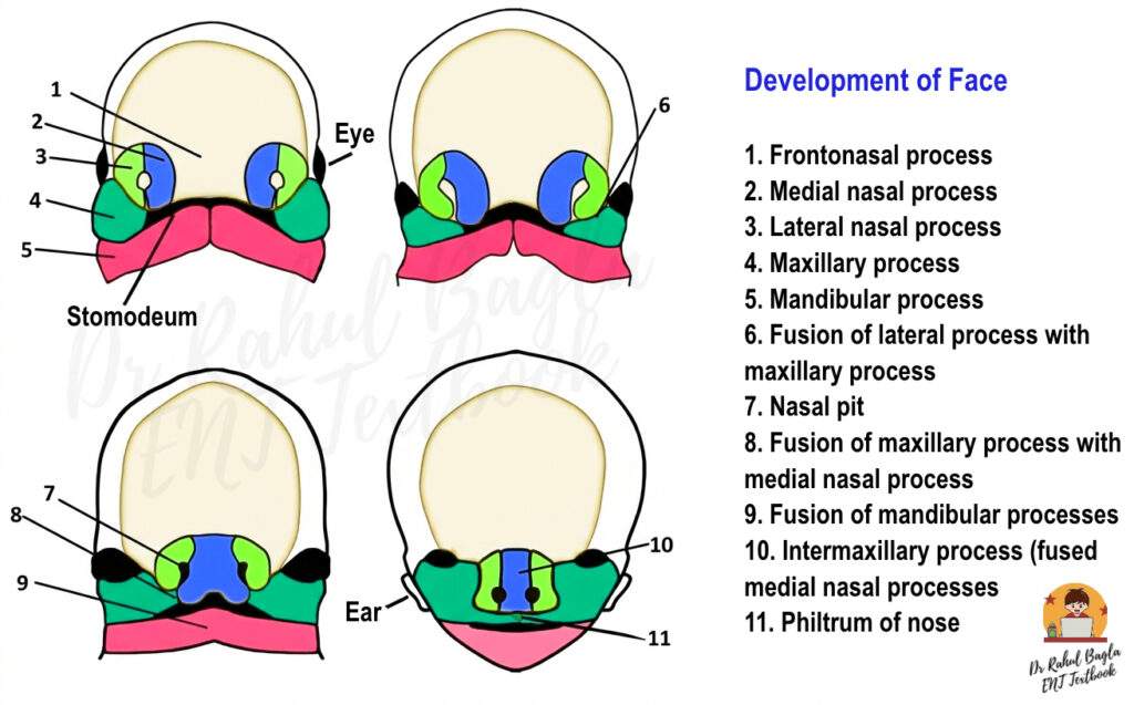
Development of the Face
Facial development begins in the 4th week of gestation. Five primordial structures, known as swellings, appear around the stomodeum (primitive mouth), which lies below the developing brain. These swellings form the basic framework of the face.
The Five Facial Primordia are:
- Frontonasal Process: Forms the forehead and the bridge of the nose.
- Right & Left Maxillary Processes: Form the cheeks and upper lip.
- Right & Left Mandibular Processes: Form the lower jaw.
All nasal structures develop through interaction between the frontonasal and maxillary processes.
Development of the External Nose and Nasal Cavities
- Stage 1: Formation of Nasal Placodes (Week 4–5). Ectoderm on the frontonasal process thickens to form nasal placodes. These placodes lie superior and lateral to the stomodeum. These oval placodes develop into the nose. They represent the earliest sign of nasal development.
- Stage 2: Formation of Nasal Pits (Week 5–6). By the 5th week, mesenchyme around each placode proliferates and grows. This growth pushes the placodes inward and forms depressions, known as nasal pits.
- Stage 3: Formation of Nasal Sacs and Primitive Choanae (Week 6). The nasal pits deepen to form nasal sacs, which grow toward the oral cavity. A thin buccanasal membrane separates the nasal and oral cavities at this stage. This membrane later ruptures to form the primitive choanae. Clinical correlation: Failure of this membrane to rupture leads to choanal atresia.
- Stage 4: Formation of Medial and Lateral Nasal Processes. Mesoderm around the nasal sacs differentiates into medial and lateral nasal processes. These processes shape the nostrils (nares). The medial nasal process forms the septum, philtrum, and primary palate. The lateral nasal process forms the alae of the nose.
- Stage 5: Fusion with Maxillary Process (Week 6–7). By the end of 6th weeks, maxillary processes grow medially and fuse with the nasal processes and frontonasal process This fusion forms the separate nasal cavities. At the junction of the lateral nasal and maxillary processes, ectoderm thickens to form epithelial cords that later canalize into the nasolacrimal duct and lacrimal sac. The lateral nasal prominence forms the nasal bones, upper lateral cartilages, and the lateral crus of the lower lateral cartilage.
| Flowchart |
| Frontonasal Process → Nasal Placodes (Week 4) → Nasal Pits → Nasal Sacs → (Membrane rupture) Primitive Choanae → Fusion with Maxillary Process → Definitive Nasal Cavity + Palate |
Development of the Upper Lip and Palate
Formation of the Upper Lip
Between the 7th and 8th weeks, the two medial nasal processes fuse and the maxillary processes. Consequently, this fusion forms the central part of the upper lip (philtrum) and the primary palate. However, if this fusion fails, a cleft lip occurs. Therefore, the mesenchyme must penetrate these junctions to ensure a solid union of the upper jaw.
Formation of the Palate: Primary vs. Secondary
The palate originates from two distinct embryological sources:
- Primary Palate: Formed by the fusion of the nasomedial processes. It eventually becomes the premaxilla, which holds the upper incisor teeth.
- Secondary Palate: Formed by the palatal shelves (outgrowths of the maxillary processes). Initially, these shelves grow vertically alongside the tongue. However, as the mandible grows and the tongue drops, the shelves flip horizontally and fuse in the midline.
Clinical Note: The incisive foramen serves as the landmark dividing the primary and secondary palate and their partial fusion leads to submucous cleft palate and non-union leads to cleft palate.
Development of the Paranasal Sinuses (PNS)
The sinuses develop as diverticula of the nasal mucous membrane. While some are present at birth, others appear much later.
Table : Summary of Sinus Development
| Sinus | Appearance (Gestation/Age) | Radiologically Visible | Adult Size Reached |
| Maxillary | 7–10 Weeks (Gestation) | At birth | 17–18 Years |
| Ethmoid | 9–10 Weeks (Gestation) | At birth | 12 Years |
| Sphenoid | 12 Weeks (Gestation) | 3 Years | 18 Years |
| Frontal | 16 Weeks (Gestation) | 8 Years | 18 Years |
Maxillary Sinus
This is the first sinus to appear. It begins as a shallow groove in the ethmoidal infundibulum. Although it is small at birth, it undergoes rapid expansion during the first seven years of life.
Ethmoid Sinus
The lateral nasal wall develops folds called ethmoturbinals. Consequently, these folds give rise to specific structures:
- 1st Ethmoturbinal: Agger nasi and Uncinate process.
- 2nd Ethmoturbinal: Bulla ethmoidalis.
- 3rd Ethmoturbinal: Basal lamella of the middle turbinate (Crucial for FESS surgery).
- 4th & 5th Ethmoturbinals: Superior and supreme turbinates.
Sphenoid Sinus
The sphenoid sinus originates from the sphenoethmoidal recess. Furthermore, clinicians categorize its pneumatization into three types relative to the sella turcica:
- Sellar (90%): Most common; pneumatization extends behind the sella.
- Pre-sellar (9%): Pneumatization reaches the anterior wall of the sella.
- Conchal (1%): Minimal pneumatization; makes transsphenoidal surgery difficult.
Frontal Sinus
The frontal sinus is the most variable. Because it originates from the anterior ethmoid cells, it only becomes a distinct “frontal” cavity after birth. It is clinically “silent” on X-rays until age 8.
Table: Important Clinical Correlations
| Developmental Error | Clinical Condition |
| Bucconasal membrane persistence | Choanal atresia |
| Maxillary–medial nasal fusion failure | Cleft lip |
| Palatal shelf fusion failure | Cleft palate |
| Ethmoid overdevelopment | Concha bullosa |
| Abnormal sphenoid pneumatization | Pituitary surgery risk |
———— End of the chapter ————
High-Yield Points for Quick Revision
- Nose develops from frontonasal process.
- Choanal atresia = failure of bucconasal membrane rupture.
- Maxillary sinus is first to develop.
- Ethmoid sinus present at birth.
- Frontal sinus absent in neonates.
- Sellar pneumatization most common (90%).
- Incisive foramen marks primary-secondary palate junction.
- Nasolacrimal duct forms from ectodermal cord between maxillary and lateral nasal processes.
Download the full PDF Link:
Reference Textbooks.
- Scott-Brown, Textbook of Otorhinolaryngology-Head and Neck Surgery.
- Cummings, Otolaryngology-Head and Neck Surgery.
- Stell and Maran’s, Textbook of Head and Neck Surgery and Oncology.
- Ballenger’s, Otorhinolaryngology Head And Neck Surgery
- Susan Standring, Gray’s Anatomy.
- Frank H. Netter, Atlas of Human Anatomy.
- B.D. Chaurasiya, Human Anatomy.
- P L Dhingra, Textbook of Diseases of Ear, Nose and Throat.
- Hazarika P, Textbook of Ear Nose Throat And Head Neck Surgery Clinical Practical.
- Mohan Bansal, Textbook of Diseases of Ear, Nose and Throat Head and Neck Surgery.
- Hans Behrbohm, Textbook of Ear, Nose, and Throat Diseases With Head and Neck Surgery.
- Logan Turner, Textbook of Diseases of The Nose, Throat and Ear Head And Neck Surgery.
- Arnold, U. Ganzer, Textbook of Otorhinolaryngology, Head and Neck Surgery.
- Ganong’s Review of Medical Physiology.
- Guyton & Hall Textbook of Medical Physiology.
Author:

Dr. Rahul Bagla
MBBS (MAMC, Delhi) MS ENT (UCMS, Delhi)
Fellow Rhinoplasty & Facial Plastic Surgery.
Renowned Teaching Faculty
Mail: msrahulbagla@gmail.com
India
———– Follow us on social media ————
- Follow our Facebook page: https://www.facebook.com/Dr.Rahul.Bagla.UCMS
- Follow our Instagram page: https://www.instagram.com/dr.rahulbagla/
- Subscribe to our Youtube channel: https://www.youtube.com/@Drrahulbagla
- Please read. Anatomy of External Ear. https://www.entlecture.com/anatomy-of-ear/
- Please read. Anatomy of Temporal Bone. https://www.entlecture.com/anatomy-of-temporal-bone/
- Please read. Stenger’s, Chimani Moos, Teal test. https://www.entlecture.com/special-tuning-fork-tests/
Keywords: Simplified embryology for quick revision.✔ Nasal placode ✔ Medial & lateral processes ✔ Cleft anomaliesPerfect for viva & theory exams. Embryology Of Nose And Paranasal Sinuses Notes For MBBS, Embryology Of Nose Summary For NEET PG, Development Of Nasal Cavity Easy Explanation, Paranasal Sinus Development Timeline Chart, Choanal Atresia Embryology Viva Questions, CBME ENT Embryology Notes PDF, Nasal Placode Development Explanation, Maxillary Sinus Development Age And Growth, Ethmoid Sinus Present At Birth Concept, Frontal Sinus Development Radiology Age, Sphenoid Sinus Pneumatization Types Exam, ENT Embryology Mnemonics For Revision, Development Of Palate And Nasal Septum Notes, Clinical Embryology Of Nose MCQs With Answers, NEET PG High Yield ENT Embryology, Applied Embryology For FESS Surgery Understanding, Lateral Nasal Wall Development Turbinates Explanation, Medical Students ENT Embryology Exam Guide