
|
The following CBME core competencies are covered in this chapter.
|
Deep Neck Space Infections: Anatomy, Clinical Features, Diagnosis and Treatment
Introduction
Deep neck space infections are infections involving the potential fascial spaces of the neck. They include retropharyngeal, danger, prevertebral, carotid, submandibular, parapharyngeal, peritonsillar, parotid, masticator and visceral space infections. These infections can spread rapidly along fascial planes and may cause airway obstruction, sepsis, vascular complications and descending mediastinitis.
Understanding cervical fascia and the communication between deep neck spaces is essential for predicting the route of infection, interpreting CT findings and planning surgical drainage. The most clinically important pathway is the relationship between the retropharyngeal space, danger space and mediastinum, which explains how an infection in the neck can descend into the thorax. This chapter provides a complete exam-oriented and clinical review of deep neck space infections, including cervical fascia, classification of neck spaces, boundaries, contents, sources of infection, clinical features, investigations, treatment, complications and important surgical relationships.
Before understanding head and neck space infections, we need to understand how the cervical fascia forms the potential spaces of the neck. The cervical fascia, first described by Burns in 1811, consists of connective tissue that ranges from loose areolar tissue to dense fibrous bands. It surrounds the muscles, nerves, blood vessels, lymphatics and viscera of the neck and forms fascial planes and potential spaces that compartmentalise the neck. These fascial planes can direct or limit the spread of infection and provide important surgical dissection planes.
Understanding the cervical fascia and neck spaces helps explain the spread of deep neck space infections, airway compromise, malignancy and their complications. Surgeons also use these fascial planes to identify important structures and perform safer dissections.
Core concept: Fascial layers → potential spaces → communication between spaces → pathways of disease spread → clinical manifestations
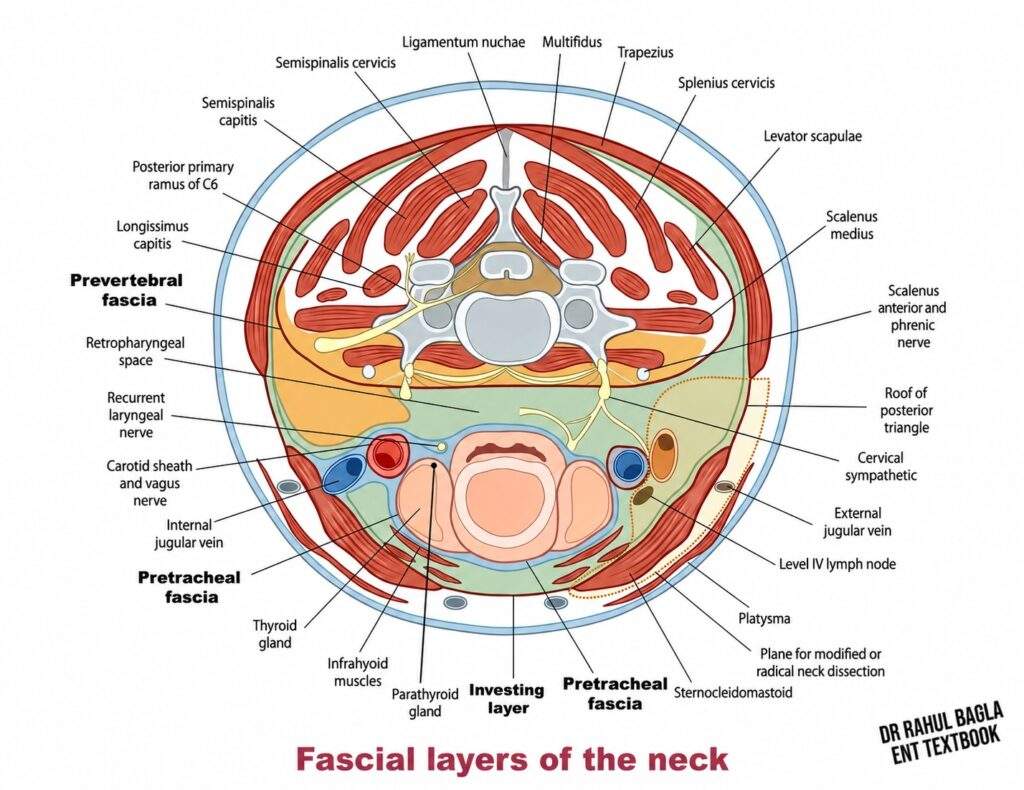
Anatomy of Cervical fascia
The cervical fascia can be broadly divided into:
- Superficial cervical fascia
- Deep cervical fascia
Superficial cervical fascia
The superficial cervical fascia lies between the dermis and the investing layer of deep cervical fascia. It resembles the subcutaneous tissue of other parts of the body. It encloses the platysma, superficial veins, including the external jugular vein, cutaneous nerves, superficial lymphatics, superficial cervical lymph nodes, loose areolar tissue and variable amounts of fat. The superficial cervical fascia allows the skin to move over deeper structures. It also provides an important plane for raising subplatysmal skin flaps during neck surgery. The external jugular vein runs within this superficial fascial plane and pierces the investing fascia before joining the subclavian vein. Therefore, surgeons should recognise it during procedures involving the lateral neck.
Clinical Importance: Infection in the superficial fascial plane usually produces cellulitis or a localised abscess. However, infection can extend into deeper neck spaces through anatomical communications or breaches in fascial boundaries.
Deep cervical fascia
The deep cervical fascia consists of dense connective tissue layers that surround and compartmentalise the deeper structures of the neck.
For practical learning, describe the deep cervical fascia as three main layers:
| Layer | Main structures enclosed or invested | Important clinical relationship |
| Investing layer (Superficial layer) | Sternocleidomastoid and trapezius; contributes to fascial coverings of the parotid and submandibular glands | Roof of posterior triangle and surgical planes |
| Pretracheal layer (Middle layer) | Infrahyoid muscles and cervical viscera | Visceral compartments and extension toward the superior mediastinum |
| Prevertebral layer (Deep layer) | Vertebral column, prevertebral muscles (longus capitis, longus colli), scalene muscles, and levator scapulae. | Posterior triangle floor; prevertebral and danger spaces |
The carotid sheath represents an important fascial compartment associated with the deep cervical fascia. It contains the major cervical neurovascular structures.
1. Investing layer of deep cervical fascia
The investing layer forms the most superficial part of the deep cervical fascia and surrounds the neck like a cylindrical fascial sleeve. It encloses the sternocleidomastoid and trapezius muscles and forms the roof of the posterior triangle of the neck between them.
Posteriorly, it attaches to the cervical spinous processes and ligamentum nuchae. Superiorly, it attaches to the external occipital protuberance, superior nuchal line, mastoid region and mandible. Inferiorly, it attaches to the clavicle, manubrium, acromion process and spine of the scapula
Relationship with the Parotid and Submandibular Glands: The investing fascia divides around the parotid gland to form the parotid sheath. Medially, the fascial covering is relatively thin and is related to the parapharyngeal space. It also surrounds the submandibular gland and forms the stylomandibular ligament, which extends from the styloid process to the angle of the mandible.
Suprasternal Space of Burns: Above the manubrium, the investing fascia splits into superficial and deep layers, creating the suprasternal space of Burns. This small potential space contains loose connective tissue and the jugular venous arch.
2. Pretracheal layer of deep cervical fascia
The pretracheal layer, traditionally called the middle layer of deep cervical fascia, mainly occupies the anterior neck. It has muscular and visceral components. The muscular component surrounds the infrahyoid strap muscles: sternohyoid, omohyoid, sternothyroid and thyrohyoid. The visceral component surrounds the thyroid gland, larynx, trachea and cervical oesophagus.
Superiorly, it attaches to the hyoid bone. Inferiorly, it extends into the superior mediastinum and blends with the fibrous pericardium. Laterally, it connects with the carotid sheath. Posteriorly, the visceral component continues as the buccopharyngeal fascia, which covers the pharyngeal constrictors and forms the anterior boundary of the retropharyngeal space. These fascial relationships help explain the spread of deep neck space infections from the pharynx toward the mediastinum.
Remember: In the simplified conventional model:
Pharyngeal wall → buccopharyngeal fascia → retropharyngeal space → alar fascia → danger space → prevertebral fascia.
This sequence is more useful clinically than memorising isolated fascial names.
3. Prevertebral layer of deep cervical fascia
The prevertebral fascia forms the deepest part of the deep cervical fascia. It covers the vertebral column and surrounds the deep muscles of the neck, including the longus capitis, longus colli, scalene muscles and levator scapulae. Superiorly, it attaches to the skull base and extends downward toward the thoracic inlet, where it continues with the endothoracic fascia.
The fascia closely covers the vertebral bodies and extends along the vertebral column. Laterally, it covers the scalene muscles, brachial plexus and subclavian vessels and forms the floor of the posterior triangle of the neck.
Alar Fascia: The alar fascia lies anterior to the prevertebral fascia and extends from the skull base to approximately T2. It separates the retropharyngeal space anteriorly from the danger space posteriorly. The alar fascia forms a fascial boundary between the retropharyngeal space and the danger space. Therefore, if infection reaches the danger space, it can extend inferiorly into the posterior mediastinum and cause potentially life-threatening mediastinitis.
Relation to Brachial Plexus: The brachial plexus and subclavian artery pass between the anterior and middle scalene muscles. The subclavian vein passes anterior to the anterior scalene muscle. Fascial extensions accompany the brachial plexus and subclavian vessels toward the axilla and contribute to the axillary sheath.
Carotid sheath
The carotid sheath is a specialized fascial sheath extending from the skull base toward the thoracic inlet. Its principal contents are the common carotid artery, internal carotid artery, internal jugular vein and vagus nerve (CN X). The vagus nerve usually lies posteriorly between the artery and vein. Deep cervical lymph nodes are closely associated with the carotid sheath and carotid space but are not considered principal contents. The sympathetic trunk lies posterior and medial to the sheath.
Several cranial nerves have important relationships with the upper carotid sheath and carotid space, including CN IX, CN XI and CN XII; however, they are not principal contents of the carotid sheath. The vagus nerve (CN X) is the principal cranial nerve within the sheath.
Traditionally, the carotid sheath is described as receiving contributions from the investing, pretracheal and prevertebral fasciae; however, its precise fascial origin remains controversial. Because it forms a longitudinal pathway through the neck, the carotid space is clinically important for spread of infection, head and neck malignancy and surgical dissection.
How Cervical Fascia Forms Neck Spaces
The cervical fascia does more than cover the structures of the neck. It creates fascial planes and potential spaces between muscles, vessels, nerves and viscera. These spaces normally contain only loose connective tissue. However, infection, pus, blood, fluid or tumour can collect within them and make the spaces clinically apparent.
Understanding these spaces helps explain the pathways of deep neck space infections and their complications. For easier learning, classify the important neck spaces according to their relationship with the hyoid bone and their anatomical pathways of spread.
Why Are Deep Neck Spaces Clinically Important?
Deep neck spaces normally contain loose connective tissue and allow neck structures to move. However, infections, bleeding and tumours can involve these spaces. Because several spaces communicate with each other, infection can spread beyond its original site. Understanding the fascial boundaries helps predict the direction of spread. It also explains the clinical findings, CT appearances and complications of deep neck infections.
How Does Infection Spread Between Neck Spaces?
Infection usually follows the path of least resistance along fascial planes. For example, a pharyngeal infection can enter the retropharyngeal space and spread into the danger space. It can then descend toward the mediastinum. Similarly, infection in the parapharyngeal space can spread to the retropharyngeal, carotid or masticator spaces. Therefore, identifying the involved space helps predict the structures at risk and possible complications.

Classification of Neck Spaces
1. Spaces Extending Along the Length of the Neck
- Retropharyngeal space
- Danger space
- Prevertebral space
- Carotid space
2. Suprahyoid Spaces
- Submandibular space – Sublingual space & Submaxillary space
- Parapharyngeal space
- Peritonsillar space
- Parotid space
- Masticator space
- Temporal space
3. Infrahyoid Spaces
- Visceral space
- Suprasternal space
Table: Deep Neck Space Infections: High-Yield Comparison
Use the table for rapid revision before studying individual spaces. First, identify the location of each space. Then link it to its symptoms, clinical findings and complications. For exams, focus on the key clinical clue that distinguishes each space. This approach is better than memorising isolated anatomical boundaries.
| S. No. | Space | Extent & key anatomical feature | Symptoms | Clinical features | Treatment |
| 1 | Retropharyngeal | Skull base → upper thoracic region; lies behind the pharynx and communicates with parapharyngeal and danger spaces | Fever, throat/neck pain, dysphagia, odynophagia | Posterior pharyngeal wall bulge, neck stiffness, torticollis, stridor | IV antibiotics + drainage for large abscesses, failed medical therapy, or airway compromise |
| 2 | Danger | Skull base → diaphragm; lies between alar and prevertebral fascia | Fever, neck pain, chest pain, respiratory difficulty | Descending mediastinitis, systemic toxicity | IV broad-spectrum antibiotics + urgent drainage; thoracic drainage if mediastinal extension |
| 3 | Prevertebral | Skull base → coccyx; related to vertebral column and deep neck muscles | Neck pain, stiffness, dysphagia, fever | Midline pharyngeal bulge, neurological deficits | Cause-specific treatment; ATT for TB, antibiotics for pyogenic infection, drainage when indicated |
| 4 | Carotid | Skull base → thoracic inlet; contains carotid arteries, IJV and vagus nerve | Fever, neck pain, swelling, dysphagia | IJV thrombosis, cranial nerve palsies, Horner syndrome | IV antibiotics + drainage of significant abscess; manage vascular complications |
| 5 | Submandibular | Around submandibular gland, below floor of mouth; divided by mylohyoid into sublingual and submaxillary compartments | Floor-of-mouth swelling, dysphagia, odynophagia | Tongue elevation, drooling, trismus, airway compromise | Secure airway + IV antibiotics + drainage when abscess is present; treat dental source |
| 6 | Parapharyngeal | Skull base → hyoid; inverted cone divided into prestyloid and poststyloid compartments | Fever, severe sore throat, odynophagia, dysphagia | Trismus, pharyngeal/tonsillar bulge, cranial nerve palsies | IV antibiotics + surgical drainage for significant abscess or complications |
| 7 | Peritonsillar | Around palatine tonsil; between tonsillar capsule and superior constrictor | Severe unilateral throat pain, fever, odynophagia | Hot-potato voice, uvular deviation, trismus | Antibiotics + aspiration or incision and drainage; tonsillectomy in selected recurrent cases |
| 8 | Parotid | Skull base → angle of mandible; enclosed by investing fascia with a thin medial fascia | Painful parotid swelling, fever, facial pain | Pus from Stensen duct, parotid tenderness | Hydration + antibiotics + drainage of significant abscess; treat predisposing factors |
| 9 | Masticator | Skull base → angle of mandible; contains muscles of mastication and V3 | Jaw pain, facial swelling, fever | Severe trismus, mandibular tenderness | IV antibiotics + drainage of significant abscess; treat odontogenic source |
| 10 | Temporal | Temporal region → zygomatic arch/infratemporal region; surrounds temporalis in superficial and deep compartments | Temporal pain, swelling, fever | Temporal swelling and trismus | IV antibiotics + surgical drainage when abscess is significant; treat dental source |
| 11 | Visceral | Skull base → superior mediastinum; surrounds pharynx, oesophagus, larynx and trachea | Dysphagia, odynophagia, neck pain, hoarseness | Respiratory difficulty, mediastinal spread, sepsis | Airway management + IV antibiotics + drainage; thoracic drainage if mediastinal extension |
| 12 | Suprasternal | Immediately above manubrium; contains loose tissue and jugular venous arch | Usually asymptomatic | Potential bleeding during tracheostomy | Usually no specific treatment; control bleeding if injured |
Retropharyngeal Space
Anatomy: The retropharyngeal space, also called the posterior visceral, retrovisceral or retroesophageal space, lies behind the pharynx and cervical oesophagus. It extends from the skull base to the upper thoracic region and continues inferiorly toward the posterior mediastinum, with the exact inferior extent varying between T1 and T6.
Boundaries:
- Anterior: Buccopharyngeal fascia behind the pharyngeal constrictors
- Posterior: Alar fascial component of the prevertebral fascia
- Lateral: Communicates with the parapharyngeal space, particularly above the hyoid bone
- Midline: A fibrous raphe divides it into right and left compartments, traditionally called the spaces of Gillette
Contents: The retropharyngeal space contains loose areolar tissue and retropharyngeal lymph nodes (nodes of Rouvière). These lymph nodes are prominent in young children and usually regress by about 3–5 years of age. In adults, retropharyngeal lymphadenopathy on imaging should raise suspicion of metastatic head and neck malignancy, particularly from pharyngeal tumours.
Source of Retropharyngeal Space Infection: Infection can reach the retropharyngeal space from the nasopharynx, adenoids, paranasal sinuses, middle ear, eustachian tube, tonsils, or parapharyngeal space. Direct infection can also follow trauma, foreign-body injury or perforation of the pharynx or cervical oesophagus. A collection of pus in the retropharyngeal space is called a retropharyngeal abscess.
Acute Retropharyngeal Abscess
Aetiology: Acute retropharyngeal abscess occurs mainly in young children, particularly those younger than 3 years, because their retropharyngeal lymph nodes are more developed. It commonly results from suppuration of retropharyngeal lymph nodes following an upper respiratory tract infection.
Clinical Features:
- Child looks sick and toxic
- Fever and malaise
- Severe throat and neck pain
- Odynophagia and dysphagia
- Trismus
- Inability to swallow saliva, causing drooling
- Neck stiffness and torticollis, with the head tilted to one side
- Stridor due to airway obstruction
- A smooth, usually unilateral bulge of the posterior pharyngeal wall
A large abscess can push the posterior pharyngeal wall and airway anteriorly, causing potentially life-threatening airway obstruction.
Key distinguishing feature: The posterior pharyngeal wall bulge is typically unilateral because the midline fibrous raphe divides the retropharyngeal space into two compartments. In contrast, a prevertebral abscess usually produces a central or midline bulge.
Diagnosis:
- Contrast-enhanced CT of the neck: This is the preferred investigation. It can distinguish phlegmon from abscess, demonstrate a rim-enhancing fluid collection, define the extent of infection and identify complications such as mediastinal extension.
- X-ray soft tissue neck: It may show widening or bulging of the prevertebral soft tissue. Normal prevertebral space is <7 mm at C2 and <14 mm at C6 in children and <7 mm at C2 and <22 mm at C6 in adults. A width of >30 mm at C6 suggests an abscess. As a general rule, widening of the prevertebral space by more than half the body size of the corresponding vertebra should raise suspicion of a retropharyngeal abscess.
- Ultrasound: May guide aspiration in selected cases.
- Laboratory studies: CBC, CRP and blood cultures.
Treatment:
1. Airway assessment: Airway assessment is the first priority. Patients with significant airway compromise require urgent airway control. Awake fibreoptic intubation may be appropriate in a difficult or compromised airway, while tracheostomy may be required when intubation is not possible or fails.
2. Antibiotics:
- Start immediate broad-spectrum IV antibiotics, such as ampicillin-sulbactam (50 mg/kg every 6 hours) or a third-generation cephalosporin with metronidazole.
- Clindamycin (15 mg/kg every 8 hours) may be used as an alternative in selected patients.
- Add vancomycin or linezolid when MRSA coverage is indicated.
- Dexamethasone may be considered as an adjunct to reduce inflammation and airway oedema.
- Treatment usually continues for 2–3 weeks, with longer treatment for complicated infections.
3. Surgical drainage: Drainage is indicated for a large or well-formed abscess, failure of medical treatment, or airway compromise. Small abscesses <2 cm, without airway compromise or severe symptoms, may be managed with antibiotics alone. A commonly used threshold is >2–2.5 cm, although clinical findings and response to treatment should guide the decision.
- Transoral drainage: Suitable for an abscess limited to the retropharyngeal space; a vertical incision is made in the posterior pharyngeal wall. The patient should be intubated and placed in the Trendelenburg position during drainage of a retropharyngeal abscess.
- External drainage: Considered for a large abscess with significant parapharyngeal extension; a transcervical approach may be required. Retract the sternocleidomastoid muscle and great vessels posteriorly to expose the retropharyngeal space. Explore the space digitally and break down all loculations, then irrigate the wound thoroughly. Place drains at appropriate sites to maintain postoperative drainage. Carefully assess patients with parapharyngeal or retropharyngeal abscesses for tracheostomy when airway compromise is present.
- Positioning: Rose position, with the head extended and slightly lowered.
- Anaesthesia: General anaesthesia with appropriate airway protection. Awake fibreoptic intubation may be required in a difficult or compromised airway, with tracheostomy as a backup when necessary.
Chronic Retropharyngeal Abscess
Aetiology: A chronic retropharyngeal abscess usually results from tuberculosis involving the cervical spine or retropharyngeal lymph nodes. It is more commonly seen in adults when associated with cervical spine disease.
Types:
- Lateral type: Usually results from tuberculous involvement of the retropharyngeal lymph nodes. It presents as a cold abscess with minimal inflammatory signs and may occur in children, particularly those younger than 5 years. It produces a lateral pharyngeal bulge.
- Central type: Usually results from Pott’s spine. The abscess develops between the vertebrae and prevertebral fascia and is therefore called a prevertebral abscess. It produces a midline, non-tender bulge and may cause neck pain and restricted neck movement.
Investigations:
- Lymphocytosis, raised ESR and positive Mantoux test
- Lateral X-ray of the cervical spine: May show caries spine, increased retropharyngeal shadow and loss of cervical lordosis
- CT: Defines the site and size of the abscess and demonstrates associated spinal involvement
Treatment:
- Antitubercular therapy
- Drainage is generally avoided unless the patient develops significant respiratory distress or dysphagia.
- Aspiration or drainage may be performed after appropriate orthopaedic consultation, particularly when there is significant spinal involvement or a large symptomatic collection.
Complications and Clinical Significance
Important complications of retropharyngeal infection include:
- Airway obstruction
- Laryngeal oedema
- Spread into the danger space
- Descending mediastinitis, which is potentially life-threatening
The retropharyngeal space lies immediately anterior to the alar fascia. Infection can therefore enter the danger space, which contains loose areolar tissue and extends inferiorly toward the thorax. Although normally a potential space, infection can fill it with fluid or pus and make it visible on imaging. Infection can then descend from the neck into the mediastinum and cause life-threatening mediastinitis.

Danger Space
Anatomy: The danger space, also called the space of Grodinsky, lies posterior to the retropharyngeal space. It extends from the skull base to the diaphragm.
Boundaries:
- Anterior: Alar fascia
- Posterior: Prevertebral fascia
The danger space is named for its direct communication with the mediastinum to the diaphragm. The danger space contains loose areolar tissue, which offers little resistance to the spread of infection. Therefore, infection from the retropharyngeal space can enter the danger space and descend into the posterior mediastinum. This can result in severe mediastinal infection and potentially life-threatening mediastinitis.
Clinical features: Infection usually reaches the danger space from an adjacent deep neck infection, particularly following rupture or extension of a retropharyngeal abscess. Once infection enters the danger space, it can descend rapidly towards the thorax. A patient with a deep neck infection who develops chest pain, respiratory difficulty or systemic toxicity should raise suspicion of mediastinal extension.
Diagnosis
- Contrast-enhanced CT of the neck and chest is the key investigation. It identifies the location and extent of infection and can demonstrate gas, fluid collections, fascial-plane involvement and mediastinal extension.
- MRI: Provides superior soft-tissue contrast and can help evaluate fascial-plane involvement in selected cases.
- Laboratory studies: CBC, CRP and blood cultures help assess systemic infection and guide treatment.
Treatment: Danger-space infection requires urgent, aggressive multidisciplinary management because of the risk of descending mediastinitis.
1. Airway: Assess and secure the airway when necessary.
2. Antibiotics: Start broad-spectrum IV antibiotics covering aerobic and anaerobic organisms, with MRSA coverage when indicated. Prolonged treatment, often 3–6 weeks, may be required when mediastinal infection is present.
3. Surgical drainage: Prompt drainage of infected collections is essential.
- Cervical approach: Used when infection remains confined to the neck.
- Cervico-thoracic approach or VATS: May be required when infection extends into the mediastinum.
- Image-guided drainage: May be appropriate for selected accessible collections.
4. Supportive care: Patients with severe infection may require ICU monitoring, haemodynamic support and management of respiratory or multiorgan complications.
Complications: The most serious complication is descending necrotising mediastinitis, which can rapidly become life-threatening. Other complications include:
- Purulent pericarditis
- Empyema
- Sepsis and septic shock
- Acute respiratory distress syndrome (ARDS)
- Carotid artery erosion with massive haemorrhage
Clinical significance: The danger space provides a low-resistance pathway for infection to travel from the deep neck into the thorax. Therefore, early recognition, appropriate antibiotics, airway management and timely surgical drainage are essential to reduce morbidity and mortality.
Prevertebral Space
Anatomy: The prevertebral space lies deep to the prevertebral fascia and extends along the vertebral column from the skull base to the coccyx. It contains the vertebral column and paraspinal musculature.
Boundaries
- Anterior: Prevertebral fascia
- Lateral: Fascial attachments to the transverse processes
- Posterior: Anterior longitudinal ligament, vertebral bodies and deep paraspinal muscles
The dense fascial attachments of this space help localise infection and limit its spread along the neck.
Aetiology: Infection of the prevertebral space most commonly results from tuberculosis of the cervical spine (spinal caries), which can produce a Pott’s abscess. Other causes include penetrating trauma, traumatic perforation of the pharynx or cervical oesophagus, and breach of the prevertebral fascia by an adjacent deep neck infection.
Clinical Features: Patients with prevertebral space infection commonly present with neck pain and stiffness, fever, dysphagia and torticollis. Neurological symptoms such as radiculopathy, myelopathy or limb weakness may occur when the infection causes nerve-root or spinal cord compression. Weight loss and night sweats suggest a tuberculous aetiology. A prevertebral abscess typically produces a central or midline bulge of the posterior pharyngeal wall, whereas a retropharyngeal abscess more commonly produces a unilateral bulge.
Diagnosis
- Contrast-enhanced CT of the neck: Demonstrates fluid collections, vertebral destruction and the extent of infection.
- MRI with contrast: Provides better assessment of spinal cord compression, epidural abscess and discitis/osteomyelitis.
- Laboratory investigations: CBC, CRP and ESR.
- Tuberculosis work-up: Interferon-gamma release assay or Mantoux test, as appropriate.
- CT-guided fine-needle aspiration or bone biopsy: Provides material for microbiological and pathological diagnosis when required.
Treatment: Treatment depends on the underlying cause.
Tuberculous abscess:
- Antitubercular therapy (ATT): A standard four-drug regimen with isoniazid, rifampicin, pyrazinamide and ethambutol, followed by continuation therapy according to the clinical response and local guidelines.
- Surgical drainage: Consider for large or symptomatic abscesses, neurological compromise or failure of medical treatment.
- Spinal stabilisation: Required when there is significant vertebral destruction or spinal instability.
Pyogenic abscess:
- Broad-spectrum IV antibiotics, with MRSA coverage when indicated.
- Surgical drainage for large or clinically significant abscesses or when medical treatment fails.
Complications: Epidural abscess, Vertebral osteomyelitis, Spinal instability, Spinal cord compression with neurological deficit, Meningitis, Sepsis, and Pott’s paraplegia in tuberculous disease
Clinical significance: Prevertebral infection is important because it can involve the vertebrae, intervertebral discs, epidural space and spinal cord. Early imaging, particularly MRI, is important when neurological symptoms or spinal involvement is suspected.
Carotid Sheath Space (Visceral Vascular Space)
Anatomy: The carotid sheath space, also called the carotid or visceral vascular space, is a potential space within the carotid sheath. The investing, pretracheal and prevertebral layers of deep cervical fascia contribute to the carotid sheath. It extends from the skull base to the thoracic inlet and continues toward the mediastinum.
Contents: The space contains the common and internal carotid arteries, internal jugular vein and vagus nerve (CN X). The sympathetic trunk and deep cervical lymph nodes lie closely related to it. Because it contains little loose connective tissue, infection usually spreads less readily through this space than through the retropharyngeal or danger spaces. The carotid space has traditionally been called “Lincoln’s highway” because it can provide a longitudinal route for infection from the neck toward the mediastinum. Infection commonly spreads into it from the parapharyngeal space.
Clinical Features: Patients may present with fever, neck pain, swelling along the anterior border of the sternocleidomastoid, torticollis and dysphagia. Trismus may occur with parapharyngeal involvement. Neurovascular involvement can cause hoarseness (vagus nerve), Horner’s syndrome (sympathetic chain), CN IX–XII palsies, or internal jugular vein thrombophlebitis, which may present as a tender, indurated cord along the sternocleidomastoid. Severe infection may cause sepsis.
Diagnosis: Contrast-enhanced CT of the neck is the preferred investigation and can demonstrate carotid sheath inflammation, abscess formation, internal jugular vein thrombosis, gas and mediastinal extension. Ultrasound with Doppler is useful for detecting internal jugular vein thrombosis and assessing vascular flow. MRI may provide better assessment of neurovascular and cranial nerve involvement. CBC, CRP and blood cultures help assess systemic infection and identify the causative organism.
Treatment: Airway assessment is the priority. Start broad-spectrum IV antibiotics covering aerobic and anaerobic organisms, with MRSA coverage when indicated. Treatment usually continues for 2–3 weeks and may require 4–6 weeks for complicated infections. Drain large abscesses, collections that fail to respond to medical treatment, or those causing complications. External drainage is usually performed along the anterior border of the sternocleidomastoid, with careful protection of the carotid artery, internal jugular vein and vagus nerve. Internal jugular vein thrombosis may require anticoagulation, although its use remains controversial and should be individualised. Carotid blowout requires urgent surgical or endovascular management and carries a mortality of >50% despite intervention. Severe infection or mediastinal extension may require ICU care.
Lemierre syndrome classically presents with pharyngitis, internal jugular vein thrombosis and septic emboli, usually to the lungs. Fusobacterium necrophorum is the commonest causative organism, particularly in young adults. Contrast-enhanced CT demonstrates internal jugular vein thrombosis, while blood cultures help identify the pathogen. Treatment requires prolonged IV antibiotics; anticoagulation remains controversial.
Complications: Important complications include internal jugular vein thrombophlebitis (Lemierre syndrome), carotid blowout, cranial nerve palsies, Horner’s syndrome, mediastinitis, sepsis and septic shock.
Submandibular Space
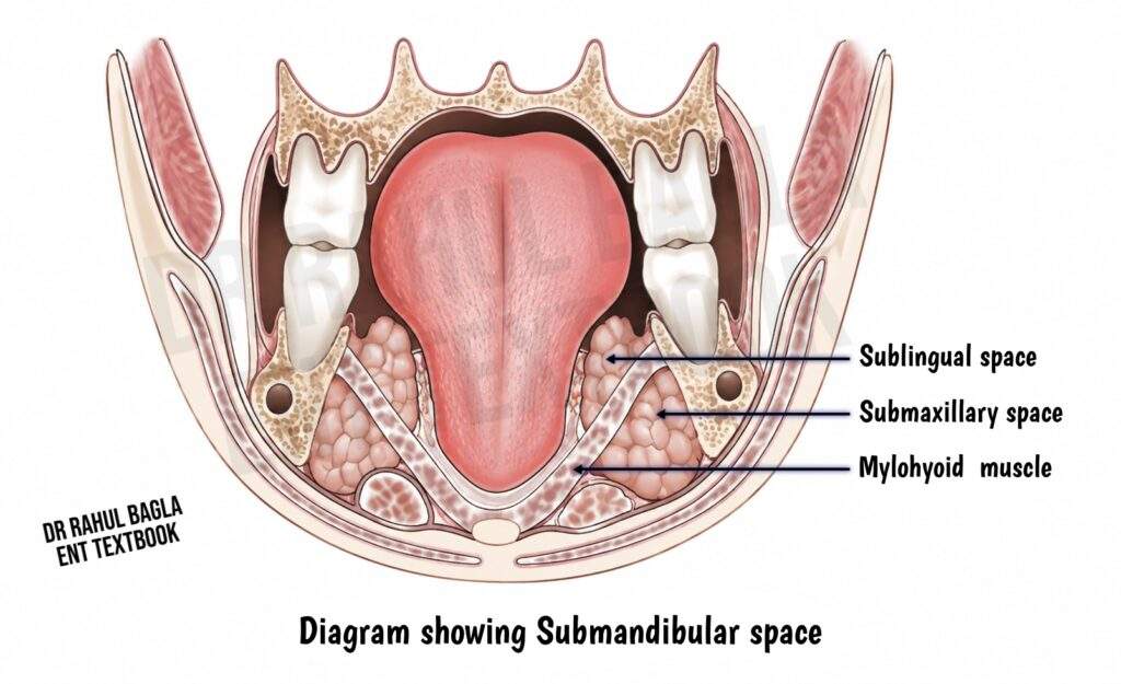
Anatomy: The submandibular space lies below the floor of the mouth and around the submandibular gland.
Boundaries:
- Superior: Lower border of the mandible
- Anterior: Anterior belly of the digastric muscle
- Posterior: Posterior belly of the digastric muscle and stylohyoid muscle
- Floor: Mylohyoid (anteriorly) and hyoglossus (posteriorly)
The mylohyoid muscle divides the submandibular space into two communicating compartments:
- Sublingual space (supramylohyoid): lies above the mylohyoid and contains loose areolar tissue, the sublingual gland, lingual nerve (CN V3) and Wharton’s duct.
- Submaxillary space (submylohyoid): lies below the mylohyoid and contains the submandibular gland, hypoglossal nerve (CN XII), facial artery, facial vein and anterior bellies of the digastric muscle.
These compartments communicate freely around the posterior border of the mylohyoid. This communication facilitates the spread of infection between the floor of the mouth and the submandibular region. The submandibular region also contains level Ib lymph nodes and the marginal mandibular branch of the facial nerve. The facial vein lies superficial to the submandibular gland, while the facial artery lies deep to the gland.
Ludwig’s Angina
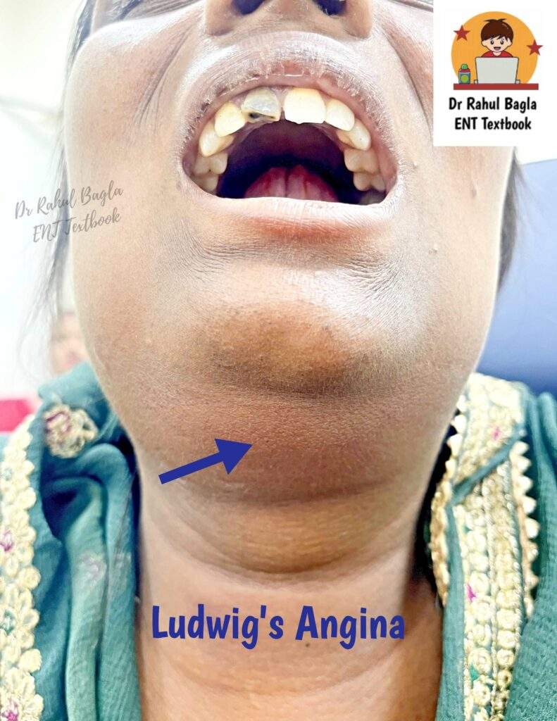
Definition: A severe form of submandibular space infection causing rapidly spreading cellulitis of the floor of the mouth, with marked swelling and induration. It was first described by Wilhelm Friedrich von Ludwig in 1836.
Criteria for Diagnosis
- It begins with rapidly spreading cellulitis without any abscess formation.
- It spreads directly along fascial planes and not by lymphatic spread.
- There is no involvement of the submandibular gland or lymph nodes.
- It involves the submaxillary and sublingual spaces and is usually bilateral.
Predisposing factors: The majority of patients will have dental caries or recent dental treatment, with diabetes mellitus or immunocompromised states like AIDS or organ transplantation.
Aetiology: Dental infection is the most common cause of submandibular space infection. Infections from the lower second and third molars commonly spread below the mylohyoid line and involve the submaxillary space. This is because the roots of the second and third molars penetrate the mylohyoid ridge, and infection of these can cause submaxillary space involvement. Infections from the premolars more often spread above the mylohyoid line into the sublingual space. Other causes include submandibular sialadenitis, penetrating injury to the floor of the mouth, mandibular fractures, tonsillar infections, foreign bodies, lymphadenitis and neoplasms.

Causative Organisms: Streptococcus viridans, Fusobacterium nucleatum, Peptostreptococcus and Actinomyces
When infection involves the sublingual space, the swelling elevates and pushes the tongue backwards. This reduces the available airway and can rapidly lead to respiratory obstruction. Infection involving the submandibular space produces a firm, tender, “woody” swelling of the floor of the mouth and neck. This finding reflects extensive cellulitis rather than a well-localised abscess.
Laryngeal oedema can further narrow the airway and worsen respiratory distress. Therefore, airway assessment and protection are the most important priorities in severe submandibular space infection and Ludwig’s angina.
Diagnosis: Ludwig’s angina is primarily a clinical diagnosis, and CT is not routinely required to confirm it.
- Use contrast-enhanced CT when you suspect spread to other deep neck spaces, and the patient can safely undergo imaging.
- Ultrasound can help differentiate cellulitis from an abscess and guide aspiration. CBC, CRP and blood cultures support the assessment of systemic infection.
- Dental evaluation is essential to identify and treat the source.
Treatment
- Secure the airway: Keep the patient upright and monitor for airway compromise (look for stridor, drooling, voice change, inability to lie flat). Perform awake fibreoptic intubation for a threatened airway and perform tracheostomy if intubation fails or is not possible.
- Start antibiotics: Give high-dose IV broad-spectrum antibiotics covering aerobes, anaerobes, and MRSA. Example: ampicillin-sulbactam + metronidazole + vancomycin (for MRSA coverage). Duration: 2–3 weeks minimum
- Reduce oedema: Give IV dexamethasone as an adjunct to reduce oedema and cellulitis.
- Surgical Drainage: Drainage is indicated for large abscesses, failure of medical treatment, or complications. Abscesses above the mylohyoid muscle (sublingual abscess) can usually be drained intraorally, while abscesses behind and below the mylohyoid (submaxillary abscess) usually require a transcervical incision in the submandibular skinfold, about 2–3 cm below the mandible. Care should be taken to avoid the marginal mandibular branch of the facial nerve. The abscess is opened by blunt dissection, and finger dissection is used to break all loculations. The cavity is then thoroughly irrigated, and a drain is placed.
- Decompress the submandibular spaces: Make a horizontal cervical incision when indicated. Drainage often produces watery oedema fluid rather than pus because Ludwig’s angina causes cellulitis rather than a localised abscess.
- Treat the dental source: Identify and treat the offending dental infection. Extract the involved tooth when appropriate after stabilising the patient.
- Provide supportive care: Give IV fluids and nutritional support and monitor the airway closely.
Complications: The most feared complication is airway obstruction, which remains the major cause of death. Other complications include laryngeal oedema, spread to the parapharyngeal space, descending mediastinitis, sepsis, aspiration pneumonia, internal jugular vein thrombosis and, rarely, carotid artery erosion with life-threatening haemorrhage.
Prognosis: Prompt recognition, antibiotics and appropriate airway management have greatly reduced mortality. Delayed treatment, particularly when airway compromise develops, significantly worsens the prognosis.
Parapharyngeal Space
Anatomy: The parapharyngeal space is also called the pharyngomaxillary, lateral pharyngeal or peripharyngeal space. It is an inverted cone-shaped space, with its broad base at the skull base superiorly, formed by the sphenoid and temporal bones, and its apex extending inferiorly to the lesser cornu of the hyoid bone.
Relations:
- Medial: Pharyngeal wall (superior constrictor muscle) and buccopharyngeal fascia
- Lateral: Medial pterygoid muscle, Ramus of the mandible and deep lobe of the parotid gland
- Posterior: Prevertebral fascia
- Anterior: Pterygomandibular raphe
The styloid process and its attached muscles divide the parapharyngeal space into two compartments. The division is formed by the stylopharyngeal fascia (Zuckerkandl–Testut fascia), which extends from the styloid process to the tensor veli palatini muscle and medial pterygoid plate.
- Prestyloid compartment (anterior to the styloid complex): It lies between the tonsillar fossa medially and the medial pterygoid muscle laterally. It contains fat, connective tissue, lymph nodes, the maxillary artery and branches of the mandibular nerve, including the inferior alveolar, lingual and auriculotemporal nerves. The deep lobe of the parotid gland is closely related to this compartment.
- Poststyloid compartment (posterior to the styloid complex): It lies posterolateral to the pharyngeal wall, medial to the parotid gland, and anterior to the prevertebral fascia. It contains the carotid sheath and its contents, the sympathetic trunk, upper deep cervical lymph nodes and cranial nerves IX, X, XI and XII.
The parapharyngeal space communicates with the peritonsillar, submandibular, parotid, masticator, retropharyngeal and carotid spaces. Therefore, it acts as an important pathway for the spread of deep neck space infections.
Clinical Features: Parapharyngeal space infection commonly develops from infections of the tonsils, pharynx or teeth (typically originating from the lower last molar tooth). A ruptured peritonsillar abscess can directly spread into this space. Parotid, retropharyngeal and submandibular space infections can also extend into it. Ear infections, such as mastoiditis with a subperiosteal abscess (historically called Bezold’s abscess) and petrositis, and penetrating trauma may occasionally cause infection. Penetrating injuries to the neck and complications from local anaesthetic injections for tonsillectomy or mandibular nerve block can introduce infection into the parapharyngeal space.
Clinical Presentation: Patients usually present with fever, severe sore throat, odynophagia, trismus and systemic toxicity.
- Prestyloid infection commonly causes medial displacement or bulging of the tonsil and lateral pharyngeal wall. Marked trismus occurs because inflammation involves the medial pterygoid muscle. Cranial nerves are not involved. Patients may also develop swelling behind the angle of the mandible.
- Poststyloid infection may produce swelling in the parotid region, torticollis and cranial nerve palsies. Involvement of cranial nerves IX, X, XI and XII can produce significant neurological deficits. There is minimal trismus.
Radiological and Surgical Importance: CT or MRI helps identify the involved compartment and the source of infection. The displacement of the parapharyngeal fat pad also helps localise lesions. Prestyloid lesions usually displace the carotid sheath and parapharyngeal fat pad posteromedially, whereas poststyloid lesions usually displace the parapharyngeal fat pad anteromedially. Surgical drainage of a parapharyngeal abscess can be challenging because the space contains major blood vessels and cranial nerves. Prompt antibiotics, appropriate drainage and careful airway assessment are essential.
Treatment:
- Airway management: Assess for stridor, drooling, voice change and inability to lie flat. Awake fibreoptic intubation is preferred; perform surgical tracheostomy if intubation fails. Avoid sedation without securing the airway.
- IV antibiotics: Start broad-spectrum IV antibiotics covering aerobic and anaerobic organisms, with MRSA coverage when indicated. Continue for 2–3 weeks, extending to 4–6 weeks for complications.
- Surgical drainage: Surgical drainage is indicated for large abscesses (>2–3 cm), failure of medical treatment, airway compromise, or complications. The parapharyngeal space is usually drained through a transcervical approach. A horizontal incision is made in a natural skin crease at the level of the hyoid bone, providing good exposure. The sternocleidomastoid muscle is retracted posteriorly, and the great vessels are identified and gently retracted to reach the abscess. The cavity is explored with a finger, all loculi are broken down, the wound is thoroughly irrigated, and a drain is placed. Transoral drainage of a parapharyngeal abscess is generally avoided because of poor exposure and the risk of injury to the carotid artery.
- Transoral drainage: Generally avoided because of the risk of major vascular injury. It may be considered for selected anterior compartment abscesses under appropriate imaging guidance.
- Management of complications: Anticoagulation with heparin/LMWH may be considered for 4–6 weeks for internal jugular vein thrombosis, although its use remains controversial. Carotid blowout requires urgent surgical or endovascular management (ligation, repair, or stenting).
- ICU monitoring: Patients with significant airway compromise, sepsis, haemodynamic instability or major complications require close ICU monitoring and supportive care.
Complications: An untreated parapharyngeal abscess can cause acute laryngeal oedema and airway obstruction. Infection may spread to the retropharyngeal and danger spaces and descend into the mediastinum. Other serious complications include internal jugular vein thrombophlebitis (Lemierre syndrome), septicaemia, carotid artery infection, mycotic aneurysm, carotid blowout, cranial nerve palsies, Horner’s syndrome, and septicemia.
Prognosis: Mortality is approximately 5–10% despite treatment; rising with delayed diagnosis; carotid blowout carries >50% mortality, making prompt diagnosis and treatment the most important prognostic factors.

Peritonsillar Space
Anatomy: The peritonsillar space is a potential space between the capsule of the palatine tonsil medially and the superior constrictor muscle laterally. It contains loose areolar tissue. The anterior and posterior tonsillar pillars form its anterior and posterior relations.
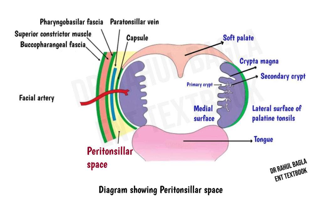
Peritonsillar Abscess: An abscess in this space is called a peritonsillar abscess, commonly known as quinsy. Peritonsillar abscess primarily affects adults, although acute tonsillitis is more common in children. The abscess is usually unilateral, though bilateral cases have been recorded. The infection is usually polymicrobial, involving both aerobic and anaerobic bacteria. Common organisms include Streptococcus pyogenes (the most common aerobic isolate), Fusobacterium necrophorum (an emerging pathogen, found in up to 22.9% of cases), Staphylococcus aureus, and anaerobes such as Prevotella and Peptostreptococcus species.
Aetiology: A peritonsillar abscess usually develops as a complication of acute tonsillitis. Infection commonly begins in a tonsillar crypt, particularly the superior tonsillar crypt (crypta magna), or from the minor salivary glands of Weber (new theory). It may spread through the tonsillar capsule into the peritonsillar space. The infection can also develop independently of acute tonsillitis. If left untreated, it may spread into the parapharyngeal space.
Clinical Features: Patients commonly present with fever, severe unilateral throat pain and odynophagia (pain in swallowing). Severe pain may prevent swallowing of saliva and cause drooling. Other common features include:
- Muffled “hot potato” voice
- Foul-smelling breath
- Trismus due to irritation of the pterygoid muscles
- Tender and enlarged jugulodigastric lymph nodes
- Ipsilateral referred ear pain due to involvement of the glossopharyngeal nerve (CN IX)
Examination: Examination typically shows swelling and congestion of the tonsil, soft palate and tonsillar pillars on the affected side. The soft palate usually bulges above and lateral to the tonsil, and the uvula becomes oedematous and is displaced to the opposite side. Mucopus may be visible over the tonsillar region. Cervical lymphadenopathy commonly involves the jugulodigastric lymph nodes. The patient may hold the head tilted toward the affected side (torticollis) because of pain and muscle spasm.
Diagnosis: The diagnosis is usually made clinically without specific investigations. OPG may identify a dental abscess, while cross-sectional imaging is indicated when complications are suspected. Pus culture is good practice but is not routinely essential, except in immunocompromised patients or recurrent quinsy.
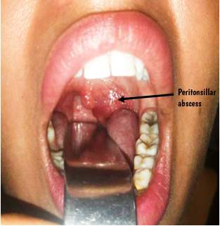
Differential Diagnosis: Parapharyngeal abscess, Odontogenic abscess (especially upper molar).
Treatment:
- Antibiotics: Early cases of peritonsillar abscess may actually represent peritonsillar cellulitis. Start antibiotics that cover Group A Streptococcus and oral anaerobes, such as penicillin, ampicillin-sulbactam, or clindamycin, usually for about 7 days. Add MRSA coverage with vancomycin or linezolid when MRSA is suspected, or the infection is severe.
- Corticosteroids: A single dose of IV dexamethasone may reduce pain, fever, trismus and hospital stay.
- Drainage: Drainage can be performed by needle aspiration using a hypodermic needle and syringe or incision and drainage (I&D). Needle aspiration is both diagnostic and therapeutic, while I&D is commonly used for a first-time abscess. Both are endorsed by AAO-HNS. The former technique is probably less reliable and may increase predisposition to re-collection of the abscess. The intraoral approach is preferred because it provides good access with low morbidity. The incision is made over the most prominent part of the swelling or at the point where a vertical line along the anterior pillar meets a horizontal line drawn at the level of the base of the uvula.
- Supportive care: Give IV fluids and analgesics and advise warm saline mouthwashes.
- Tonsillectomy (AAO-HNS 2019 Guideline): Interval tonsillectomy may be considered in patients with >1 peritonsillar abscess or repeated failed drainage, and is usually performed 4–6 weeks after the acute infection has resolved. Immediate (hot) tonsillectomy may be considered in selected patients with recurrent, bilateral, or difficult-to-drain abscesses.
Complications: An untreated peritonsillar abscess can spread into the parapharyngeal and retropharyngeal spaces. Other complications include airway obstruction, aspiration of pus causing pneumonitis or lung abscess, sepsis, Lemierre syndrome (septic thrombophlebitis of the internal jugular vein with septic emboli), and, rarely, carotid artery or jugular vein erosion causing severe haemorrhage. Intracranial complications such as brain abscess can also occur.
Parotid Space
Anatomy: The parotid space is formed by the investing layer of deep cervical fascia, which splits into superficial and deep layers and surrounds the parotid gland. The superficial layer is thick and forms a strong parotid capsule, while the deep layer is relatively thin. The thin medial fascia provides a pathway for infection to spread into the parapharyngeal space.
The parotid space contains the parotid gland, parotid lymph nodes, facial nerve, retromandibular vein and external carotid artery with its terminal branches. The facial nerve divides the parotid gland into superficial and deep surgical planes, although the gland itself does not form two separate anatomical lobes. An abscess in this parotid space is called a parotid abscess.
Aetiology: A parotid abscess is usually associated with acute bacterial infection of the parotid gland. Dehydration is an important risk factor because reduced salivary flow promotes bacterial growth. It commonly occurs in postoperative, debilitated or seriously ill patients. Bacteria can ascend from the oral cavity through Stensen’s duct and infect the parotid gland. Multiple small collections may coalesce to form an abscess. Staphylococcus aureus is the most common causative organism. Streptococci, anaerobes and occasionally Gram-negative bacteria may also cause infection.

Clinical Features: Patients usually develop painful swelling, redness and tenderness over the parotid region and angle of the mandible. The swelling is usually unilateral but may occasionally be bilateral. Fluctuation may be difficult to detect because the parotid fascia is firm. The opening of Stensen’s duct may appear congested, and gentle pressure over the gland may express pus through the duct. Fever and dehydration are common systemic features.
Differential Diagnosis:
- Acute suppurative parotitis (cellulitis without abscess)
- Viral parotitis (mumps) — bilateral, parotid enlargement, no pus
- Sialadenitis (inflammation without abscess)
- Parotid neoplasm (painless mass, no acute inflammation)
- Lymphadenitis
- Cellulitis
Diagnosis: Ultrasound can identify a parotid collection and guide aspiration. CT is useful when the abscess is deep, extensive or suspected to have spread into adjacent spaces. Aspirated pus should be sent for culture and sensitivity to guide antibiotic therapy.
Clinical Importance: The thin medial fascia of the parotid space provides a pathway for infection to spread into the parapharyngeal space. Infection can then extend into other deep neck spaces and, in severe cases, descend toward the mediastinum. Therefore, early diagnosis and treatment of a parotid abscess are important to prevent deep neck space infection and its potentially life-threatening complications.
Treatment
- Supportive care: Ensure adequate hydration, preferably with IV fluids if oral intake is poor. Encourage sialogogues such as lemon drops to promote salivary flow, maintain good oral hygiene with warm saline mouthwashes, and provide analgesics such as paracetamol or ibuprofen.
- Antibiotic therapy: Start broad-spectrum antibiotics covering S. aureus, streptococci, and anaerobes. IV ampicillin–sulbactam is commonly used; add MRSA coverage when clinically indicated. Switch to oral antibiotics once the patient improves, usually completing 7–10 days of treatment.
- Surgical drainage: Surgical drainage is considered if no improvement is seen after 24-48 hours of antibiotic therapy. Small, accessible abscesses may be treated with ultrasound-guided needle aspiration, with the pus sent for culture and sensitivity. For a large, multiloculated, or non-responding abscess, perform incision and drainage through a preauricular or facelift-type incision. The parotid capsule is opened carefully, the abscess cavity is explored and all loculations are broken down, followed by thorough irrigation and placement of a drain. The facial nerve and its branches must be identified and protected during deep dissection.
- Treat predisposing factors: Correct dehydration, optimise diabetes control, and treat underlying causes such as sialolithiasis. Recurrent or chronic infection may rarely require superficial parotidectomy.
Complications: Parotid abscess may spread to the parapharyngeal and deep neck spaces, including the masticator, retropharyngeal, and danger spaces. Infection may descend through the carotid sheath, causing mediastinitis. Other complications include facial nerve palsy, internal jugular vein thrombosis, carotid artery erosion, mandibular osteomyelitis, fistula formation, and recurrence if the underlying cause is not treated.
Masticator Space
Anatomy: The masticator space is a deep facial space enclosed by the superficial layer of deep cervical fascia. This fascia splits around the ramus of the mandible and encloses the muscles of mastication. The space extends from the angle of the mandible to the skull base, with a temporal component extending above the zygomatic arch. It lies lateral to the parapharyngeal space.
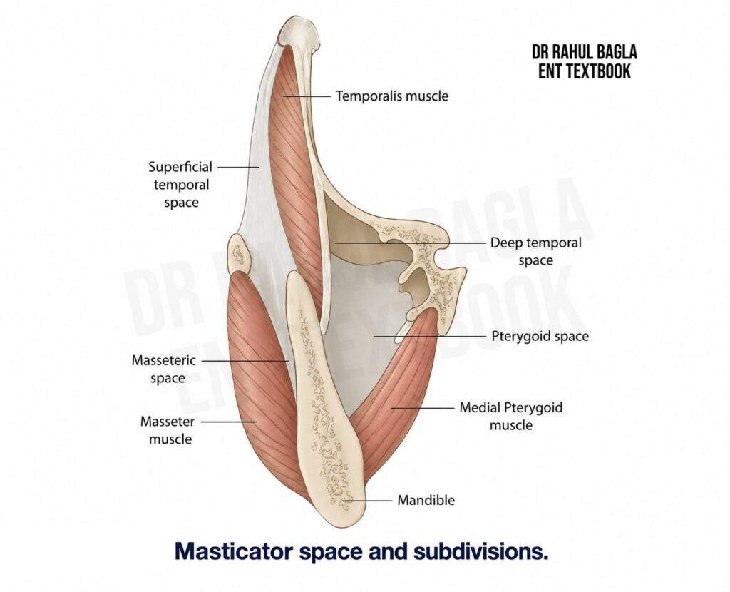
Boundaries and Relations: The mandibular ramus forms the central skeletal component of the space. The buccal space lies anteriorly, the parotid space lies posterolaterally, and the parapharyngeal space lies medially. Superiorly, the space continues into the temporal and infratemporal regions. Inferiorly, it relates to the submandibular and sublingual spaces.
Contents: The space contains the four muscles of mastication—masseter, temporalis, medial pterygoid and lateral pterygoid. It also contains the posterior body and ramus of the mandible, the mandibular division of the trigeminal nerve (V3), branches of the maxillary artery and the pterygoid venous plexus.
Compartments: The masticator space has masseteric, pterygoid and temporal components. The masseteric space lies between the masseter and mandibular ramus. The pterygomandibular component lies around the medial pterygoid and mandibular ramus. The temporal component includes the superficial temporal space, between the temporalis fascia and muscle, and the deep temporal space, between the temporalis muscle and temporal bone.
Infections: Masticator-space infection is most commonly odontogenic, particularly from infections of the mandibular molars. Infection can spread from the oral cavity or adjacent deep facial spaces and may involve multiple spaces.
Clinical Features: Trismus is the hallmark feature because infection involves the muscles of mastication. Patients may also develop facial swelling, jaw pain, fever and tenderness over the mandible.
Diagnosis: Contrast-enhanced CT (CECT) is the preferred, identifies the source of infection, demonstrates abscess formation, assesses mandibular involvement and shows extension into adjacent spaces. MRI provides better soft-tissue contrast and is particularly useful for assessing muscle involvement, marrow disease, perineural spread and intracranial extension. CBC, CRP and blood cultures help assess systemic infection.
Treatment:
- Management includes airway assessment, IV antibiotics and drainage of a significant abscess.
- Start broad-spectrum antibiotics that cover oral aerobes and anaerobes, and add MRSA coverage when clinically indicated.
- Drain large or well-formed abscesses, collections that fail to respond to medical treatment, and infections with significant complications.
- Choose an intraoral or external approach according to the location and extent of the abscess.
- Always treat the odontogenic source, including dental extraction or definitive dental treatment when indicated. Severe infections may require ICU monitoring.
Complications: Complications include mandibular osteomyelitis, spread to adjacent deep spaces, sepsis and airway compromise. Extensive infection can rarely cause intracranial complications. The masticator space also provides a potential pathway for perineural tumour spread along V3 through the foramen ovale, which is particularly important when evaluating masticator-space masses.
Temporal Space
Anatomy: The temporal space lies on the lateral aspect of the skull and surrounds the temporalis muscle. The temporalis fascia attaches superiorly to the temporal lines and inferiorly to the zygomatic arch. The temporal space contains loose connective tissue, vessels and nerves and communicates with the masticator and infratemporal spaces. The temporalis muscle divides it into superficial and deep compartments.
Boundaries: The lateral boundary is formed by the temporalis fascia, while the medial boundary is formed by the periosteum of the temporal bone. The superficial temporal compartment lies between the temporalis fascia and temporalis muscle, whereas the deep temporal compartment lies between the temporalis muscle and temporal bone.
Clinical Features: Temporal-space infection most commonly results from odontogenic infection and may spread from the adjacent masticator or infratemporal spaces. Patients may develop painful temporal swelling, fever, facial pain and trismus.
Diagnosis: Contrast-enhanced CT is the preferred investigation. It identifies cellulitis or abscess formation and demonstrates extension into the masticator and infratemporal spaces. MRI provides better soft-tissue detail and is particularly useful when intracranial extension is suspected. CBC and CRP help assess systemic infection.
Treatment: Treat temporal-space infection with broad-spectrum IV antibiotics covering oral aerobes and anaerobes, with MRSA coverage when indicated. Drain a significant or well-formed abscess surgically. During external drainage, carefully protect the temporal branch of the facial nerve. Treat the odontogenic source with appropriate dental treatment or extraction when indicated. Assess the airway in patients with extensive facial or deep neck infection.
Complications: Complications include spread to the masticator and infratemporal spaces, intracranial extension, temporal bone osteomyelitis, meningitis and sepsis. Intracranial spread may occur through skull-base pathways or venous channels.
Visceral Space
Anatomy: The visceral space is a potential space surrounding the pharynx, oesophagus, larynx and trachea. It extends from the skull base through the neck and communicates inferiorly with the superior mediastinum. The pretracheal fascia forms an important part of the fascial boundary of the anterior visceral compartment, which surrounds the cervical trachea, oesophagus and thyroid gland.
Contents: The visceral compartment contains or surrounds the pharynx, cervical oesophagus, larynx, trachea and thyroid gland. Because the fascial plane continues inferiorly toward the mediastinum, infection can spread from the neck into the chest.
Aetiology: Infection may result from perforation or injury of the cervical oesophagus, particularly after instrumentation or penetrating trauma. It may also spread from adjacent deep neck spaces or descend from infections involving the pharynx and retropharyngeal space.
Clinical Features: Patients may develop dysphagia, odynophagia, neck pain, hoarseness, respiratory difficulty and fever. Oesophageal perforation may produce subcutaneous emphysema. Severe infection can cause airway compromise and systemic sepsis.
Diagnosis: Contrast-enhanced CT of the neck and chest is the preferred investigation when deep infection or mediastinal extension is suspected. CT identifies fluid or gas collections, defines the extent of infection and demonstrates mediastinal involvement. Contrast swallow studies can help identify an oesophageal perforation when clinically suspected. CBC, CRP and blood cultures help assess systemic infection.
Treatment: First, assess and secure the airway when respiratory compromise occurs. Start broad-spectrum IV antibiotics covering aerobic and anaerobic organisms, with MRSA coverage when indicated. Keep patients with suspected oesophageal perforation or significant visceral infection nil by mouth and provide appropriate nutritional support. Drain significant collections surgically or with image guidance, depending on their location and extent. Mediastinal extension may require thoracic surgical drainage. Patients with severe infection or mediastinal involvement may require ICU monitoring.
Complications: Important complications include mediastinitis, sepsis, septic shock, pneumonia, empyema, laryngeal oedema and airway compromise.
Suprasternal Space
Anatomy: The suprasternal space, also called the space of Burns, is a small potential space located above the manubrium. It forms when the investing layer of deep cervical fascia splits into superficial and deep layers before attaching to the manubrium and clavicles.
Boundaries
- Superior: Intertendinous connection between the sternocleidomastoid muscles
- Inferior: Manubrium
- Lateral: Sternocleidomastoid muscles
- Anterior: Superficial layer of investing fascia
- Posterior: Deep layer of investing fascia
Contents: The space contains loose connective tissue and the jugular venous arch.
Clinical Significance: The suprasternal space is particularly important during tracheostomy and lower neck surgery because injury to the jugular venous arch can cause significant bleeding. The space may appear on CT or MRI as a small midline fascial space immediately above the manubrium.
Other important spaces in the head and neck are:
1. Pterygopalatine Fossa (Pterygomaxillary Space)
The pterygopalatine fossa is a pyramidal-shaped space located below the apex of the orbit. This space houses important neurovascular structures, including the maxillary division of the trigeminal nerve, the vidian nerve, the sphenopalatine nerve, the lesser and greater palatine nerves, the sphenopalatine ganglion, and the internal maxillary artery. Infections in this area typically originate from the maxillary teeth or from osteomyelitis.
- Signs and Symptoms: Gingival oedema, facial cellulitis, trismus, and ocular symptoms. Infections may also extend into the infratemporal fossa.
- Treatment: Drainage via the Caldwell-Luc approach through the oral buccal sulcus, alongside appropriate antibiotics.
2. Buccal Space
The buccal space is bounded by the buccinator muscle, the cheek, the pterygomandibular raphe, the zygomatic arch, and the inferior mandible. The most common source of infection is odontogenic in origin.
- Signs and Symptoms: Infections can cause buccal swelling that may extend to the eyelid (preseptal) and involve the orbicularis oris.
- Complications: Serious complications include cavernous sinus thrombosis, intracranial infections, and extension of the infection into other spaces and the orbit.
- Treatment: Prompt drainage, often via an external approach, and an aggressive antibiotic regimen are critical in management.
———— End of the chapter ————
High-Yield Points for NEET PG and University Exams
- Retropharyngeal abscess: posterior pharyngeal wall bulge, dysphagia, neck stiffness and torticollis.
- Danger-space infection: descending spread toward the mediastinum.
- Prevertebral infection: commonly associated with cervical spine disease and may produce a midline pharyngeal bulge.
- Ludwig’s angina: rapidly spreading bilateral cellulitis of the submandibular and sublingual spaces with potential airway obstruction.
- Parapharyngeal abscess: severe sore throat, trismus and possible cranial nerve involvement.
- Peritonsillar abscess: unilateral throat pain, hot-potato voice, trismus and uvular deviation.
- Masticator-space infection: marked trismus is a major clinical clue.
- Carotid-space infection: internal jugular vein thrombosis, cranial neuropathies and vascular complications.
NEET PG Style MCQs
- A 2-year-old child presents with fever, drooling, neck stiffness, and stridor. Examination reveals a unilateral bulge in the posterior pharyngeal wall. What is the most likely diagnosis? A. Prevertebral abscess B. Retropharyngeal abscess C. Peritonsillar abscess D. Parapharyngeal abscess.
- A patient with a deep neck infection develops chest pain, respiratory difficulty, and fever. CT shows mediastinal involvement. Which space is most likely involved? A. Retropharyngeal space B. Danger space C. Prevertebral space D. Submandibular space.
- A 45-year-old diabetic patient presents with painful submandibular swelling, tongue elevation, and stridor. What is the first priority in management? A. IV antibiotics B. Surgical drainage C. Airway assessment and protection D. CT scan.
- A patient presents with “hot potato” voice, uvula deviation to the opposite side, and trismus. What is the most likely diagnosis? A. Retropharyngeal abscess B. Parapharyngeal abscess C. Peritonsillar abscess D. Parotid abscess.
- Which space is traditionally called “Lincoln’s highway” because it provides a longitudinal route for infection spread from the neck to the mediastinum? A. Retropharyngeal space B. Danger space C. Carotid sheath space D. Prevertebral space.
- A patient with a parapharyngeal abscess develops hoarseness, Horner’s syndrome, and dysphagia. Which compartment is most likely involved? A. Prestyloid compartment B. Poststyloid compartment C. Masseteric space D. Sublingual space.
- A young adult presents with pharyngitis, tender cord along the sternocleidomastoid, and septic emboli to the lungs. What is the most likely causative organism? A. Streptococcus pyogenes B. Staphylococcus aureus C. Fusobacterium necrophorum D. Haemophilus influenzae.
- What is the hallmark feature of masticator space infection? A. Fever B. Swelling over the mandible C. Trismus D. Cranial nerve palsy.
- A patient with a parotid abscess fails to improve after 48 hours of antibiotics. What is the next best step? A. Continue antibiotics for another 48 hours B. CT scan C. Incision and drainage D. Ultrasound-guided needle aspiration.
- Which space is clinically important during tracheostomy because injury to the jugular venous arch can cause significant bleeding? A. Visceral space B. Suprasternal space of Burns C. Retropharyngeal space D. Danger space.
Answers: 1. B. 2. B. 3. C. 4. C. 5. C. 6. B. 7. C. 8. C. 9. C. 10. B.
Clinical Case Scenarios for Practical Exams and Viva
Case 1. A 3-year-old child presents with high-grade fever, drooling, neck stiffness, and stridor. The mother reports that the child has had an upper respiratory tract infection for the past 5 days. On examination, you note a unilateral bulge in the posterior pharyngeal wall.
Most likely diagnosis: Acute retropharyngeal abscess.
Best investigation: Contrast-enhanced CT of the neck.
Management: Airway assessment (awake fibreoptic intubation if compromised), IV antibiotics (ampicillin-sulbactam or third-generation cephalosporin with metronidazole), and surgical drainage if abscess >2 cm or airway compromise is present.
Case 2. A 35-year-old diabetic patient presents with rapidly progressive submandibular and submental swelling, tongue elevation, drooling, and difficulty breathing. The swelling is firm, tender, and “woody” in consistency. There is no fluctuance.
Most likely diagnosis: Ludwig’s angina.
Key distinguishing feature: Bilateral woody swelling, no abscess formation, tongue elevation, airway compromise.
Management: Secure the airway (awake fibreoptic intubation preferred), IV antibiotics (ampicillin-sulbactam + metronidazole + vancomycin for MRSA), IV dexamethasone, and surgical decompression if indicated.
Case 3. A 45-year-old patient presents with severe unilateral throat pain, fever, muffled “hot potato” voice, and drooling. Examination reveals a swollen tonsil, bulging soft palate, and uvula displaced to the opposite side.
Most likely diagnosis: Peritonsillar abscess (quinsy).
Best next step: Incision and drainage at the point of maximum bulge or at the junction of the anterior pillar and a horizontal line through the base of the uvula.
Indication for interval tonsillectomy: More than one episode of peritonsillar abscess.
Frequently Asked Questions (FAQ)
- What is the difference between retropharyngeal and prevertebral abscess? A retropharyngeal abscess produces a unilateral bulge because the midline raphe divides the retropharyngeal space into two compartments. A prevertebral abscess produces a midline bulge because it lies between the vertebrae and the prevertebral fascia.
- Why is airway management the first priority in Ludwig’s angina? Ludwig’s angina causes rapid swelling of the floor of the mouth, pushing the tongue upwards and backwards. This can cause life-threatening airway obstruction. Therefore, securing the airway is the absolute priority.
- Why is transoral drainage contraindicated in parapharyngeal abscess? The parapharyngeal space contains the carotid artery, internal jugular vein, and cranial nerves. Transoral drainage provides poor exposure and carries a high risk of injury to these structures.
- What is Lemierre syndrome?
Lemierre syndrome is septic thrombophlebitis of the internal jugular vein, most commonly caused by Fusobacterium necrophorum. It presents with pharyngitis, internal jugular vein thrombosis, and septic emboli to the lungs. It primarily affects young adults. - How do you differentiate prestyloid from poststyloid compartment infection? Prestyloid infection causes marked trismus and tonsillar displacement. Poststyloid infection causes cranial nerve palsies (IX-XII) and Horner’s syndrome with minimal trismus.
- What is the significance of the danger space? The danger space extends from the skull base to the diaphragm. It provides a low-resistance pathway for infection to spread from the neck to the mediastinum, causing life-threatening descending mediastinitis.
- When should interval tonsillectomy be considered for peritonsillar abscess? According to AAO-HNS 2019 guidelines, interval tonsillectomy should be considered in patients with more than one peritonsillar abscess or repeated failed drainage attempts. It is usually performed 4–6 weeks after the acute infection resolves.
———— End ————
Download the full PDF Link:
Head and neck space infections Best Lecture Notes Dr Rahul Bagla ENT Textbook
Reference Textbooks.
- Scott-Brown, Textbook of Otorhinolaryngology-Head and Neck Surgery.
- Cummings, Otolaryngology-Head and Neck Surgery.
- Stell and Maran’s, Textbook of Head and Neck Surgery and Oncology.
- Ballenger’s, Otorhinolaryngology Head And Neck Surgery
- Susan Standring, Gray’s Anatomy.
- Frank H. Netter, Atlas of Human Anatomy.
- B.D. Chaurasiya, Human Anatomy.
- P L Dhingra, Textbook of Diseases of Ear, Nose and Throat.
- Hazarika P, Textbook of Ear Nose Throat And Head Neck Surgery Clinical Practical.
- Mohan Bansal, Textbook of Diseases of Ear, Nose and Throat Head and Neck Surgery.
- Hans Behrbohm, Textbook of Ear, Nose, and Throat Diseases With Head and Neck Surgery.
- Logan Turner, Textbook of Diseases of The Nose, Throat and Ear Head And Neck Surgery.
- Arnold, U. Ganzer, Textbook of Otorhinolaryngology, Head and Neck Surgery.
- Ganong’s Review of Medical Physiology.
- Guyton & Hall Textbook of Medical Physiology.
Author:

Dr. Rahul Bagla
MBBS (MAMC, Delhi) MS ENT (UCMS, Delhi)
Fellow Rhinoplasty & Facial Plastic Surgery.
Renowned Teaching Faculty
Mail: msrahulbagla@gmail.com
India
———– Follow us on social media ————
- Follow our Facebook page: https://www.facebook.com/Dr.Rahul.Bagla.UCMS
- Follow our Instagram page:https://www.instagram.com/dr.rahulbagla/
- Subscribe to our Youtube channel: https://www.youtube.com/@Drrahulbagla
- Please read. Juvenile Angiofibroma. https://www.entlecture.com/juvenile-angiofibroma/
- Please read. Tumours of Hypopharynx . https://www.entlecture.com/tumours-of-the-hypopharynx/
- Please read. Anatomy of Oesophagus. https://www.entlecture.com/anatomy-of-oesophagus/
Keywords: Parotid Space, Submandibular Space, Peritonsillar Space, Retropharyngeal Space, Danger Space, Prevertebral Space, Parapharyngeal Space, Masticator Space, Head and Neck Space Infections: Causes, Signs, and Symptoms; Management of Deep Neck Space Infections; Types of Head and Neck Space Infections; Complications of Untreated Neck Space Infections; Diagnosis of Deep Neck Abscess; Surgical Treatment for Neck Space Infections; Risk Factors for Head and Neck Infections; Antibiotics for Deep Neck Infections; Prevention of Head and Neck Space Infections. Head and Neck Space Infections: Causes, Symptoms, and Treatments. Deep Neck Space Infections: Diagnosis and Management Explained. Signs and Symptoms of Head and Neck Space Infections You Shouldn’t Ignore. Complications of Untreated Head and Neck Space Infections: What to Watch For. Comprehensive Guide to Surgical and Antibiotic Treatments for Neck Infections. Understanding the Risk Factors and Prevention Strategies for Neck Space Infections. Types of Head and Neck Space Infections and How They Are Treated. Deep Neck Abscess: Causes, Diagnosis, and Effective Management. Prevention and Early Detection of Head and Neck Infections. How to Identify and Treat Severe Head and Neck Infections Safely.
Thank you sir
Nice presentation
And we need from you add
Empirical treatment for each space according
Sure.
Thanks for the appreciation.
Me encantó la presentación, muchas gracias.
Thanks