Myringotomy
It involves a surgical incision of the tympanic membrane to aspirate fluid from the middle ear. It is performed to drain suppurative or non-suppurative effusions from the middle ear cavity.
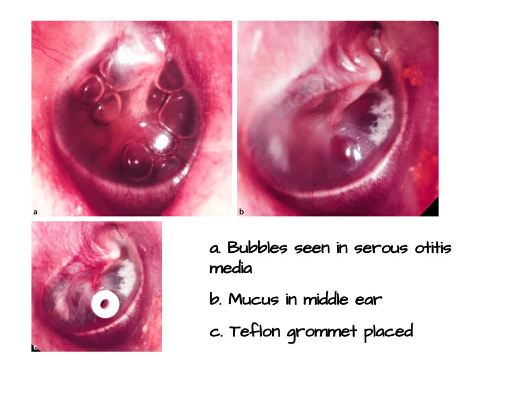
Ventilation tube (Grommet / Tympanostomy Tube)
It is also known as a grommet or pressure-equalising tympanostomy tube, which may also be placed to provide drainage or ventilation in cases of eustachian tube dysfunction. Thus, the grommet provides continuous drainage and, more importantly, ventilation to the middle ear, bypassing a dysfunctional Eustachian tube. Various materials are used for ventilation tubes, with Teflon or medical-grade silicone being most common due to their biocompatibility. Titanium, gold, or silver oxide-coated tubes are also utilised to inhibit biofilm formation.
Types of Ventilation Tubes (Based on Retention Time):
-
Short-Term Tubes (Shepard, Reuter bobbin): These spontaneously extrude after 6–18 months. They are preferred for Otitis Media with Effusion (OME).
-
Long-Term Tubes (T-tube, Paparella): These require surgical removal and can stay in place for 2–4 years. They are used for chronic or recurrent conditions like atelectasis or chronic Eustachian tube dysfunction.
Indications for Myringotomy
1. Acute suppurative otitis media
- Severe earache with a bulging eardrum about to perforate.
- Persistent tympanic membrane retraction causing conductive hearing loss
- Complications of acute otitis media, such as facial nerve paralysis, labyrinthitis, or meningitis with a bulging tympanic membrane
2. Recurrent acute otitis media: ≥ 3 episodes in 6 months or ≥ 4 episodes in 12 months.
3. Otitis media with effusion (OME)
4. Aero-otitis media (to drain fluid and “unlock” the eustachian tube)
5. Patulous eustachian tube
6. Atelectatic ear (grommet is often inserted for long-term aeration)
7. Hemotympanum
Anaesthesia for Myringotomy
General anaesthesia is routinely used in children and geriatric patients. Otherwise, myringotomy can be performed under local anaesthesia or without anaesthesia.
Surgical Technique of Myringotomy and Ventilation Tube Insertion
1. Clean the external auditory canal of wax and debris.
2. Always operate under an operating microscope.
3. The preferred site for ventilation tube insertion is the anteroinferior quadrant (right ear at 5 o’clock, left ear at 7 o’clock) through a circumferential or radial incision. Insertion of the ventilation tube in the posterosuperior quadrant is not recommended due to the risk of ossicular chain damage. For longer retention of the ventilation tube, the anterosuperior quadrant is preferred. The type of incision (radial or circumferential) does not affect extrusion rates.


4. The incision should penetrate the entire thickness of the tympanic membrane, avoiding injury to the ossicular chain, posterior meatal wall, jugular bulb, or any abnormal vascular structures (e.g., high jugular bulb, aberrant carotid artery, or glomus tympanicum).
5. Aspiration of middle ear fluid through the incision site before placing the grommet should be avoided, as it may promote biofilm infection and tympanosclerosis (secondary to trauma and bleeding of the tympanic membrane).
Postoperative Care of Myringotomy
- Topical ear drops at the time of grommet placement reduce the risk of blockage by blood or mucus and the chances of local infection during the early postoperative period.
- Avoid swimming and water entry into the ear canal as long as the grommet is in place.
- In cases of acute suppurative otitis media, perform daily cleaning of ear discharge from the canal. In serous otitis media, leave a cotton wool wick for 24-48 hours.
Complications of Myringotomy
- The most common operative complication is displacement of the ventilation tube into the middle ear. Attempts should be made to retrieve the tube, but failure to remove it rarely causes problems due to the inert nature of the grommet.
- Infection around the grommet can lead to a middle ear infection.
- Otorrhea may follow an acute upper respiratory tract infection.
- Granulation tissue may develop secondary to infection.
- Residual tympanic membrane perforation
- Pars tensa atrophy and retraction
- Blockage due to blood or secretions
- Tympanosclerosis is the most common structural complication
———— End of the chapter ————
High-Yield Points for Quick Revision (NEET PG)
- The anteroinferior quadrant (5 and 7 o’clock positions) is the standard insertion site.
- Posterosuperior quadrant insertion is contraindicated due to ossicular injury risk.
- Tympanosclerosis is the most common structural complication of grommet insertion.
- Recurrent Acute Otitis Media (RAOM) indication: ≥3 episodes in 6 months or ≥4 in 1 year.
- Otitis Media with Effusion (OME) indication: Persistent for ≥3 months with significant hearing loss.
- T-tubes (Long-term) have a higher rate of residual perforation than Shepard tubes (Short-term).
Clinical Scenarios and Viva Voce
Scenario 1: A 4-year-old child presents with a 4-month history of inattentiveness at school and frequent “misunderstanding” of speech. Pure Tone Audiometry (PTA) confirms bilateral conductive hearing loss of 35 dB. Otoscopy shows dull, thick, retracted TMs with fluid levels.
Viva Questions and Answers:
- Q: What is the diagnosis and the management? A: The diagnosis is Bilateral Chronic Otitis Media with Effusion (OME), as the effusion has persisted for ≥3 months with significant hearing loss. The definitive management is Bilateral Myringotomy and Grommet (Shepard tube) insertion.
- Q: Why do you prefer a grommet over a simple myringotomy? A: Simple myringotomy incision closes within days, and the effusion will likely recur due to persistent Eustachian tube dysfunction. The grommet provides long-term pressure equalisation and ventilation, allowing the middle ear mucosa to heal and the Eustachian tube to mature.
- Q: How long will the grommet stay in place? A: We use a short-term tube (e.g., Shepard) for OME. It typically spontaneously extrudes within 6 to 18 months.
Scenario 2: A 6-year-old who had grommets placed 3 months ago for RAOM is brought to the clinic with persistent ear discharge (otorrhea) for 3 days following a common cold.
Viva Questions and Answers:
- Q: What is the likely cause of the discharge, and how will you treat it? A: The discharge is likely acute otitis media or otitis externa that developed secondary to contamination via the grommet, often triggered by the URTI. Treatment: Start with topical antibiotic drops (e.g., quinolone-based drops, as they are non-ototoxic and safe with a grommet). We avoid oral antibiotics unless the child is toxic or the discharge persists.
- Q: What advice must you give the parents regarding water entry? A: They must prevent water entry (bathing, swimming) into the ear canal as long as the grommet is in place, as water is a major source of contamination.
Multiple Choice Questions (MCQs) for NEET PG
- The preferred site for ventilation tube insertion in the tympanic membrane is the: A. Posterosuperior quadrant B. Anterosuperior quadrant C. Anteroinferior quadrant D. Posteroinferior quadrant
- Which of the following complications is considered the most common structural change following grommet insertion? A. Residual TM perforation B. Granuloma formation C. Cholesteatoma D. Tympanosclerosis
- A child presenting with persistent Otitis Media with Effusion (OME) and significant hearing loss should be considered for grommet insertion if the effusion persists for at least: A. 6 weeks B. 2 months C. 3 months D. 6 months
- Grommet insertion is contraindicated in the posterosuperior quadrant primarily due to the risk of injury to the: A. Jugular bulb B. Facial nerve C. Ossicular chain D. Eustachian tube orifice
- Which type of ventilation tube is typically used for long-term middle ear ventilation (2–4 years) and usually requires surgical removal? A. Shepard tube B. Reuter bobbin C. T-tube D. Collar button
- For a patient with acute suppurative otitis media and a severely bulging, unruptured tympanic membrane, the primary goal of myringotomy is: A. To prevent tympanosclerosis B. To improve hearing immediately C. To drain pus and relieve pain D. To allow long-term aeration
- A patient with recurrent acute otitis media is defined by having: A. ≥2 episodes in 12 months B. ≥3 episodes in 12 months C. ≥3 episodes in 6 months or ≥4 episodes in 12 months D. ≥5 episodes in 2 years
- Which of the following structures is least likely to be injured during myringotomy in the anteroinferior quadrant? A. Annulus B. Stapes C. Handle of malleus D. Jugular bulb
- Which statement regarding fluid management during myringotomy is currently considered best practice? A. Aspirate all fluid to ensure complete drainage. B. Avoid excessive aspiration of fluid before tube placement. C. Only aspirate mucoid fluid, not serous fluid. D. Aspirate using a large-bore needle.
- The most common complication of the grommet tube function is: A. Extrusion B. Otorrhea C. Tympanosclerosis D. Residual perforation
Answers and Explanations:
- C. The anteroinferior quadrant is relatively safe and avascular.
- D. Tympanosclerosis (white patches on TM) is the most frequent structural change resulting from inflammation and trauma.
- C. The standard guideline for OME requiring intervention (grommet) is ≥3 months with documented hearing loss.
- C. The incudostapedial joint and the stapes are located in the posterosuperior quadrant.
- C. T-tubes and Paparella tubes are designed for long-term retention.
- C. The immediate priority is pain relief and preventing spontaneous rupture/complication by draining the tension.
- C. This is the standard definition used for surgical intervention (grommet).
- B. The stapes is located deep in the posterosuperior quadrant; the anteroinferior quadrant is the safest distance from it.
- B. Excessive aspiration can cause trauma, bleeding, and may promote biofilm infection and tympanosclerosis.
- B. Otorrhea (ear discharge) is the most common problem seen with an in-situ grommet, usually secondary to URTI or water contamination.
Frequently Asked Questions in Viva
- What is the medical term for the ventilation tube placed in the eardrum? The ventilation tube is also known as a grommet or a pressure-equalising (PE) tube.
- What is the main reason for inserting a grommet in a child? The primary reason is to treat persistent Otitis Media with Effusion (OME) and the resulting significant conductive hearing loss caused by Eustachian tube dysfunction.
- Do grommets fall out by themselves? Short-term grommets (like Shepard tubes) spontaneously extrude from the eardrum, usually within 6 to 18 months, as the TM heals itself.
- Can a child swim after grommet insertion? No, the child must avoid getting water into the ear canal while the grommet is in place to prevent infection (otorrhea) and middle ear contamination.
- What is the most serious structural risk of grommet insertion? Residual tympanic membrane perforation (a hole that doesn’t close after the tube falls out) is the most significant risk, especially with long-term T-tubes.
———–End————-
Reference Textbooks.
- Scott-Brown, Textbook of Otorhinolaryngology-Head and Neck Surgery.
- Glasscock-Shambaugh, Textbook of Surgery of the Ear.
- P L Dhingra, Textbook of Diseases of Ear, Nose and Throat.
- Hazarika P, Textbook of Ear Nose Throat And Head Neck Surgery Clinical Practical.
- Mohan Bansal, Textbook of Diseases of Ear, Nose and Throat Head and Neck Surgery
- Hans Behrbohm, Textbook of Ear, Nose, and Throat Diseases With Head and Neck Surgery.
- Salah Mansour, Middle Ear Diseases – Advances in Diagnosis and Management.
- Logan Turner, Textbook of Diseases of The Nose, Throat and Ear Head And Neck Surgery.
- Rob and smith, Textbook of Operative surgery.
- Arnold, U. Ganzer, Textbook of Otorhinolaryngology, Head and Neck Surgery.
Author:

Dr. Rahul Bagla
MBBS (MAMC, Delhi) MS ENT (UCMS, Delhi)
Fellow Rhinoplasty & Facial Plastic Surgery.
Renowned Teaching Faculty
Mail: msrahulbagla@gmail.com
India
———– Follow us on social media ————
- Follow our Facebook page: https://www.facebook.com/Dr.Rahul.Bagla.UCMS
- Follow our Instagram page: https://www.instagram.com/dr.rahulbagla/
- Subscribe to our Youtube channel: https://www.youtube.com/@Drrahulbagla
- Please read. Anatomy of External Ear. https://www.entlecture.com/anatomy-of-ear/
- Please read. Anatomy of Temporal Bone. https://www.entlecture.com/anatomy-of-temporal-bone/
- Please read. Stenger’s, Chimani Moos, Teal test. https://www.entlecture.com/special-tuning-fork-tests/
Keywords: Acute suppurative otitis media, Recurrent AOM, Otitis media with effusion, Aero-otitis media, Patulous eustachian tube, Atelectatic ear, Hemotympanum, ENT topics for NEET PG, MBBS ENT notes, ENT viva questions Myringotomy, Grommet insertion site, Myringotomy indications, Complications of grommet insertion, Otitis Media with Effusion management, Tympanosclerosis grommet, Myringotomy for OME in children, Ventilation tube insertion technique ENT, Indications of myringotomy in MBBS curriculum, Complications of tympanostomy tubes, How to perform myringotomy with grommet, NEET PG ENT myringotomy MCQ, Tympanic membrane incision site quadrant