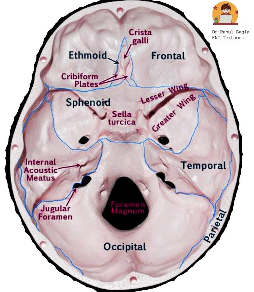
Overview of the Temporal Bone
- Paired bone forming part of lateral walls & base of the skull.
- Houses external, middle, and inner ear.
- Articulates:
- Anterior: Sphenoid bone lies between the right and left temporal bones.
- Posterior: Occipital bone
- Inferior: articulates with the Mandible → forms TMJ
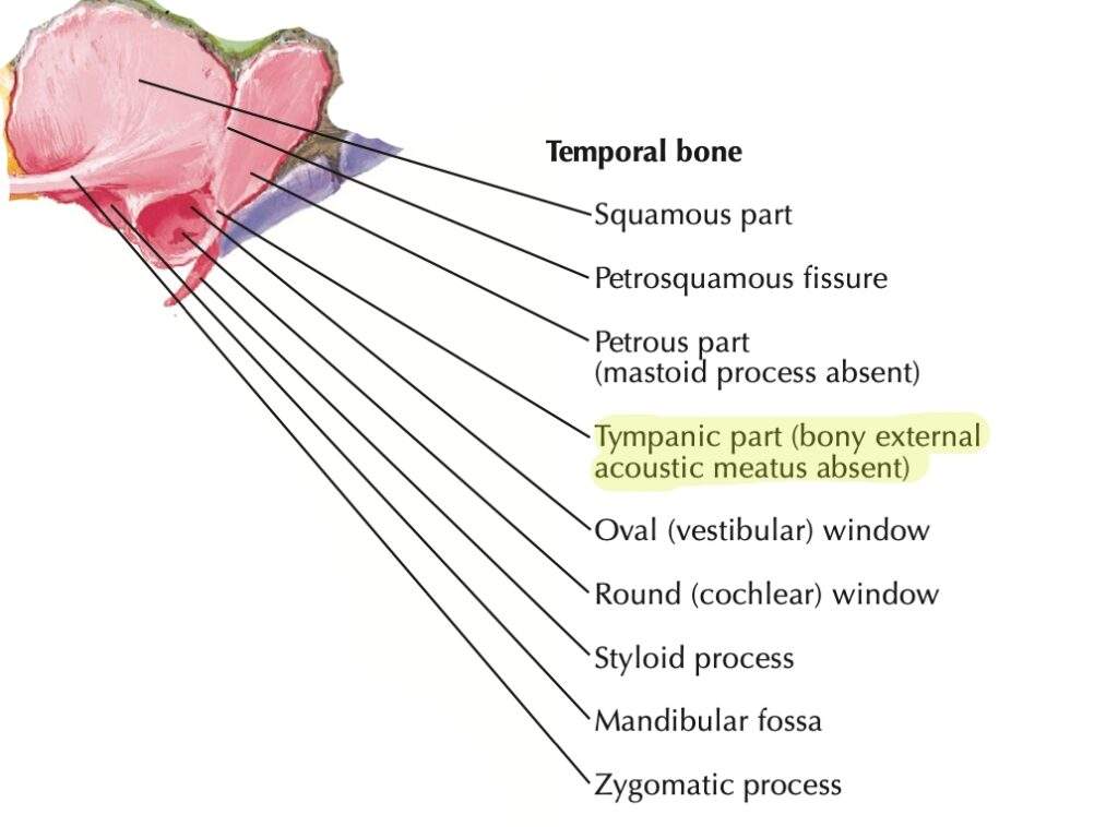
Five Parts of Temporal Bone Mnemonic: “Some People Make Thin Structures”
- Squamous
- Petrous
- Mastoid
- Tympanic
- Styloid

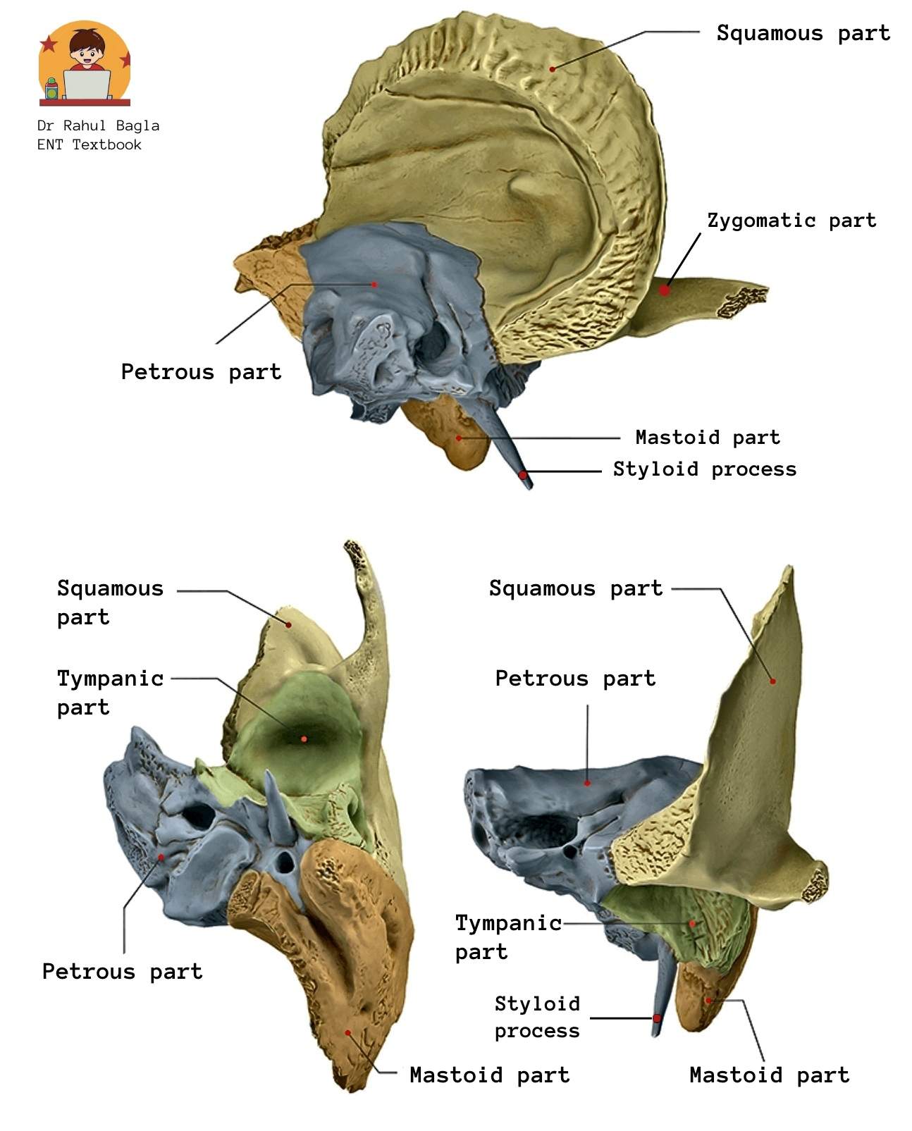
- Squamous Part
- Largest and superior most part of the temporal bone
- Flat, thin, translucent, shell-like plate of bone.
- Forms lateral wall of middle cranial fossa.
- It mainly forms the roof of the EAC and very small parts of the anterior and posterior walls of the external auditory canal (EAC).
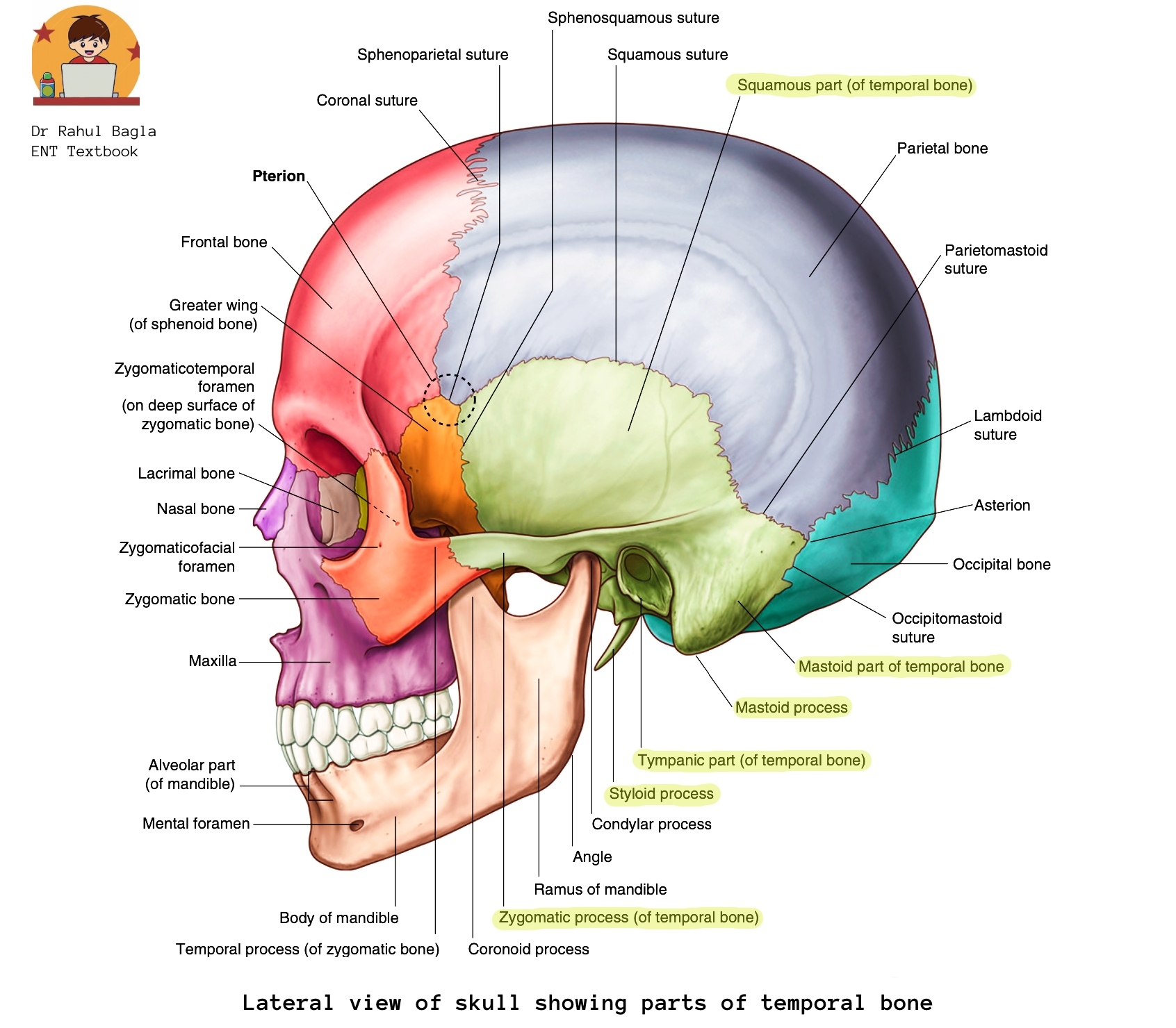
- Zygomatic process articulates with the temporal process of the zygomatic bone to form a zygomatic arch (palpable as ‘cheekbones’).
- Mandibular fossa below the zygomatic process houses the temporomandibular joint.
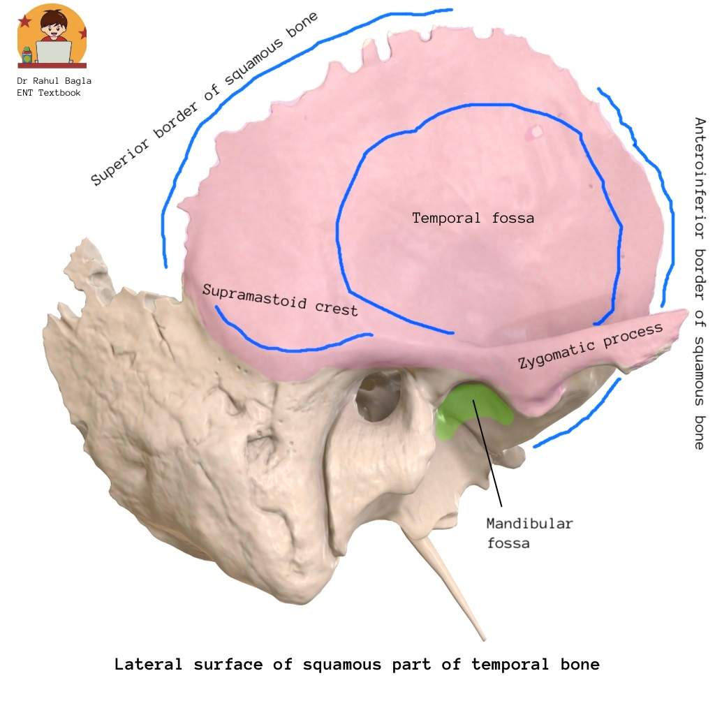
- Relations: It has two surfaces (lateral, medial) and two borders (superior, anteroinferior).
- Lateral surface forms the temporal fossa and gives origin to the temporalis muscle. Supramastoid crest on the lateral surface provides attachment to temporalis fascia.
- Medial surface is related to the temporal lobe and middle meningeal artery grooves.
- Superior border articulates with the parietal bone.
- Anteroinferior border articulates with the sphenoid.
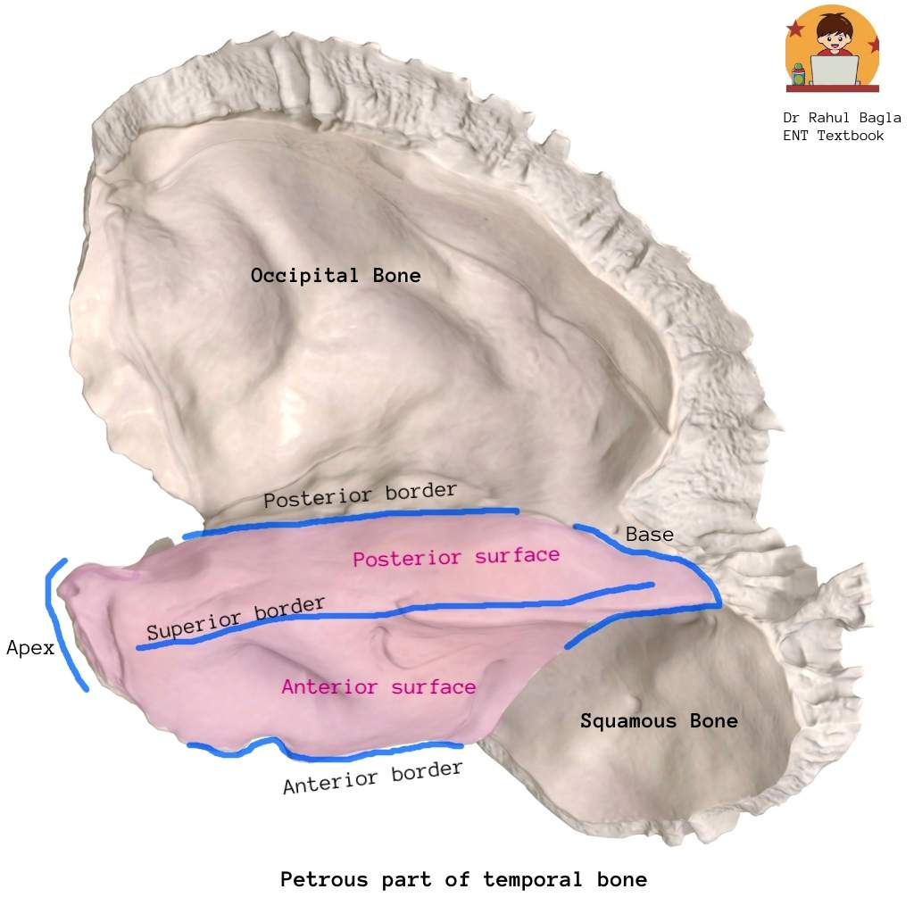
- Petrous Part (“Rock-like”)
- It is pyramidal shaped, dense bone and houses middle & inner ear, facial canal, cochlea, semi-circular canals.
- It is located in the base of the skull between the sphenoid bone anteriorly and the occipital bone posteriorly.
- Relations: It has a base, an apex, three surfaces (anterior, posterior, inferior) and three edges or margins (superior, anterior, posterior).
-
- Base: Fuses with medial surfaces of squamous and mastoid part.
- Apex: Points anteromedially; related to trigeminal (Meckel’s cave) and abducent nerves (Dorello’s canal); involved in Gradenigo’s syndrome.
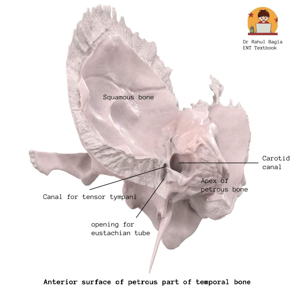
- Anterior surface: Forms posterior wall of middle cranial fossa; shows (medial to lateral) trigeminal impression, roof of internal acoustic meatus, arcuate eminence and tegmen tympani.
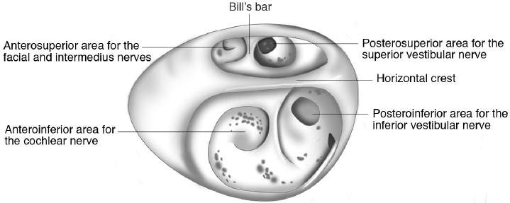
- Posterior surface: Forms anterior wall of posterior cranial fossa; contains internal acoustic meatus for facial, vestibulocochlear nerves, and artery.
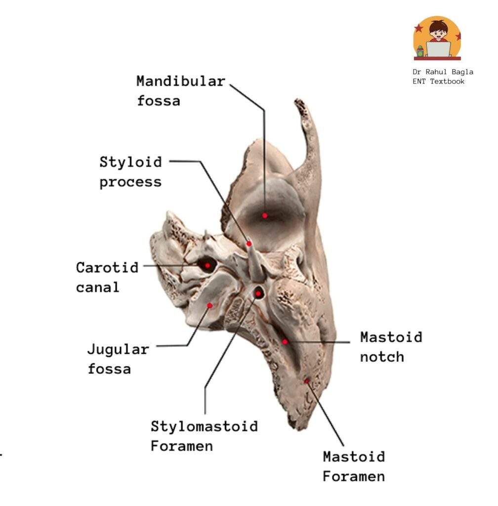
- Inferior surface: Shows carotid canal, jugular fossa, and stylomastoid foramen for facial nerve exit.
- Superior border: It is a long sharp crest between posterior and anterior cranial fossae; provides attachment for tentorium cerebelli and superior petrosal sinus.
- Anterior border: Medially joins sphenoid; laterally fuses with squamous temporal bone via petrosquamosal suture.
- Posterior border: Has groove for inferior petrosal sinus and contributes to jugular foramen boundary.

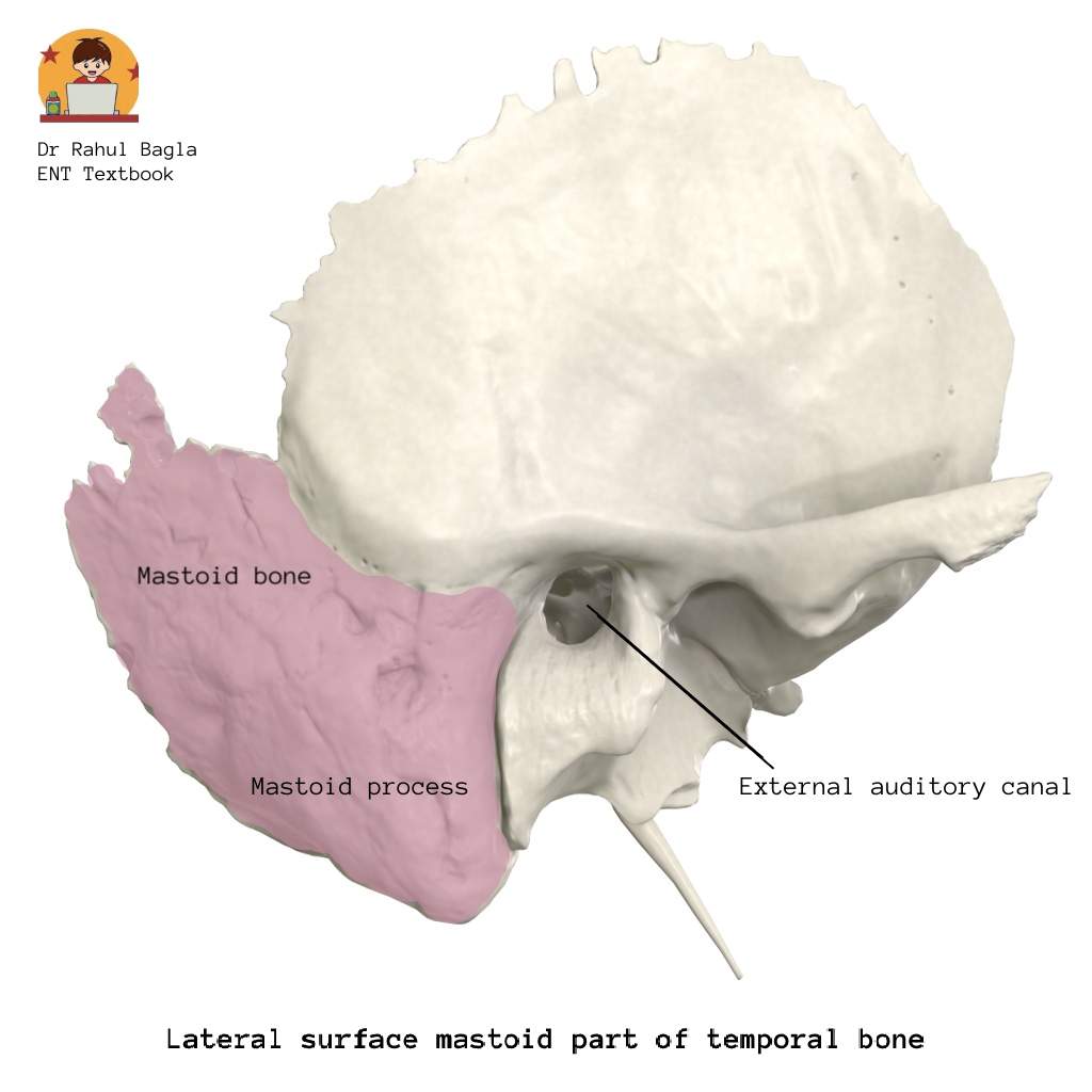
- Mastoid Part
- It lies behind the squamous part; separated by a fused suture ~1.5 cm behind the supramastoid crest.
- It contains mastoid antrum and air cells that communicate with the middle ear.
- It is formed by squamous (lateral) and petrous (medial) parts, divided by Körner’s septum.
- Relations: It has two surfaces (lateral and medial) and two borders (superior and posterior):
- Lateral surface: Forms mastoid process; gives muscle attachments (SCM, splenius, longissimus capitis).
- Medial surface: Features sigmoid sulcus which lodges sigmoid sinus, digastric notch, groove for occipital artery, and mastoid foramen for emissary vein and artery.
- Superior border forms parietomastoid suture with parietal bone.
- Posterior border joins occipital bone via occipitomastoid suture.
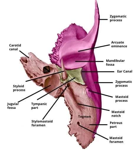
- Tympanic Part
- It lies lateral to petrous, below squamous, and in front of mastoid part of temporal bone.
- It forms anterior, posterior, and floor of bony EAC.
- Sutures: Forms tympanosquamous suture (anterior) and tympanomastoid suture (posterior).
- Petrotympanic fissure: transmits chorda tympani, anterior tympanic artery.
- Relations: It has three borders (superior, inferior & lateral) and two surfaces (anterior & posterior):
- Borders: superior (related to mandibular fossa), inferior (related to styloid process & petrous part), and lateral (forms bony ear canal)
- Anterior surface relates to parotid gland and mandibular fossa.
- Posterior surface, bears tympanic sulcus for attachment of tympanic membrane.
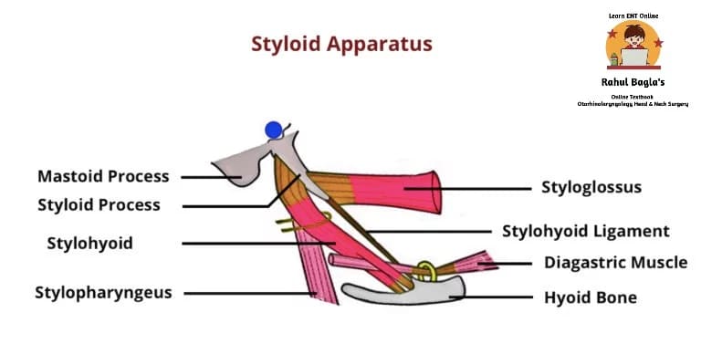
- Styloid Part (Styloid Process)
- Thin, slender, pointed bony projection which arises from the junction of tympanic and petrous parts, in front of the jugular fossa; ~2.5 cm long.
- Projects downward, forward, and medially; serves as an anchor for muscles and ligaments.
- Muscles attached: Styloglossus (CN XII), Stylohyoid (CN VII), Stylopharyngeus (CN IX).
- Ligaments attached: Stylohyoid and stylomandibular; the latter separates parotid from submandibular gland and medial pterygoid.
- Stylomastoid foramen: Lies between styloid and mastoid; transmits facial nerve (CN VII) and stylomastoid artery.
Important Foramina & Contents
| Foramen | Contents |
| Internal acoustic meatus | CN VII, VIII, labyrinthine artery |
| Stylomastoid foramen | Facial nerve (CN VII), stylomastoid artery |
| Jugular foramen | CN IX, X, XI, sigmoid sinus |
| Carotid canal | Internal carotid artery |
| Petrotympanic fissure | Chorda tympani, anterior tympanic artery |
| Mastoid foramen | Mastoid emissary vein |
———— End of the chapter ————
Reference Textbooks.
- Scott-Brown, Textbook of Otorhinolaryngology-Head and Neck Surgery.
- Glasscock-Shambaugh, Textbook of Surgery of the Ear.
- P L Dhingra, Textbook of Diseases of Ear, Nose and Throat.
- Hazarika P, Textbook of Ear Nose Throat And Head Neck Surgery Clinical Practical.
- Mohan Bansal, Textbook of Diseases of Ear, Nose and Throat Head and Neck Surgery
- Hans Behrbohm, Textbook of Ear, Nose, and Throat Diseases With Head and Neck Surgery.
- Salah Mansour, Middle Ear Diseases – Advances in Diagnosis and Management.
- Logan Turner, Textbook of Diseases of The Nose, Throat and Ear Head And Neck Surgery.
- Rob and smith, Textbook of Operative surgery.
- Anirban Biswas, Textbook of Clinical Audio-vestibulometry.
- Arnold, U. Ganzer, Textbook of Otorhinolaryngology, Head and Neck Surgery.
Author:

Dr. Rahul Bagla
MBBS (MAMC, Delhi) MS ENT (UCMS, Delhi)
Fellow Rhinoplasty & Facial Plastic Surgery.
Renowned Teaching Faculty
Mail: msrahulbagla@gmail.com
India
———– Follow us on social media ————
- Follow our Facebook page: https://www.facebook.com/Dr.Rahul.Bagla.UCMS
- Follow our Instagram page: https://www.instagram.com/dr.rahulbagla/
- Subscribe to our Youtube channel: https://www.youtube.com/@Drrahulbagla
- Please read. Anatomy of External Ear. https://www.entlecture.com/anatomy-of-ear/
- Please read. Anatomy of Temporal Bone. https://www.entlecture.com/anatomy-of-temporal-bone/
- Please read. Stenger’s, Chimani Moos, Teal test. https://www.entlecture.com/special-tuning-fork-tests/